
Patient Data
Age: 20 years
Gender: Male
From the case:
Ewing sarcoma - pelvis
Download
Info
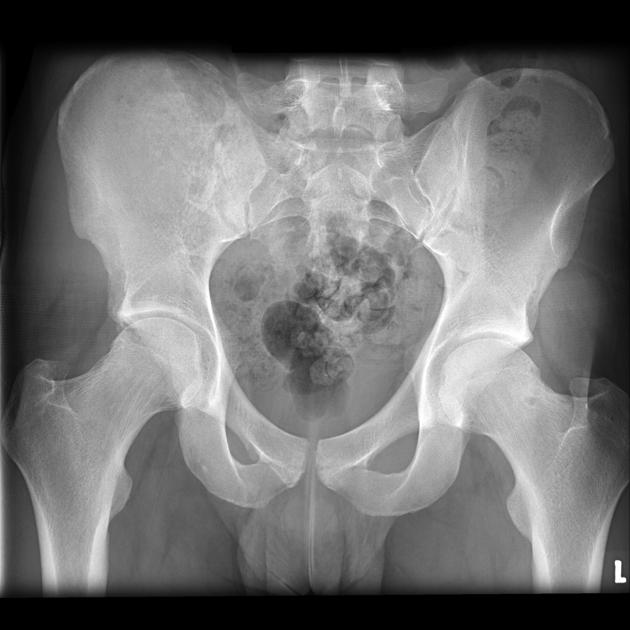
A very ill-defined lucency on the right involving the pelvis is seen, with no convincing cortical breach or periosteal reaction. There is also some soft tissue fullness lateral to the pelvis and medial displacement of the cecum.
Download
Info
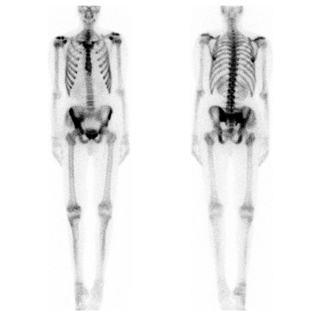
Bone scan demonstrates markedly increased uptake in the right hemipelvis.
From the case:
Ewing sarcoma - pelvis

Download
Info
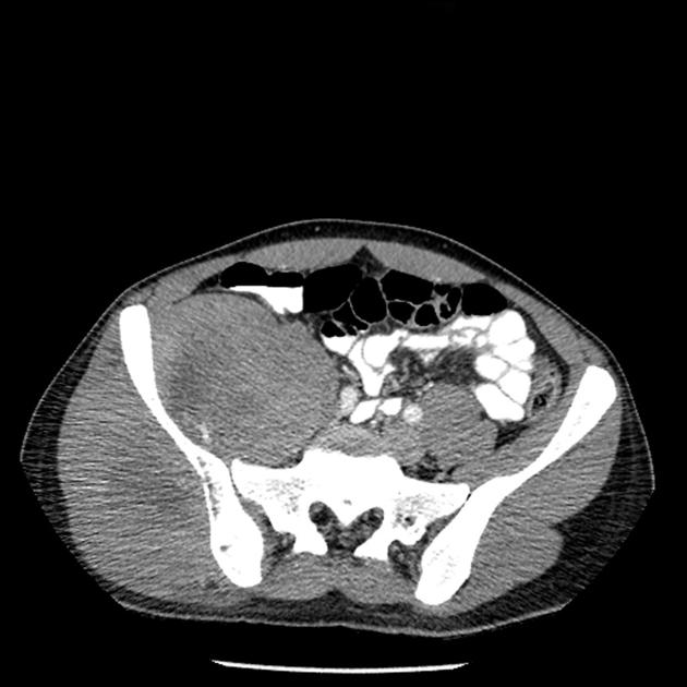
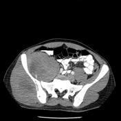
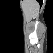
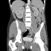
CT demonstrates a large soft tissue mass centered on the right hemi-pelvis.
From the case:
Ewing sarcoma - pelvis

Download
Info

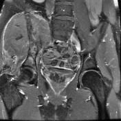
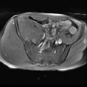
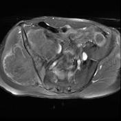
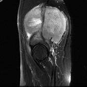
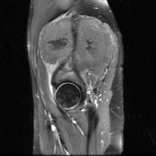
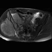
MRI confirms the CT findings, with a very large mass not evident extending through the pelvis.
- Note: This case has been tagged as "legacy" as it no longer meets image preparation and/or other case publication guidelines.
Case Discussion
This was confirmed to be a Ewing sarcoma of the pelvis in a 20 year old male.
Case courtesy of Bob Cook, MD. Western Memorial Regional Hospital Corner Brook, Newfoundland.


 Unable to process the form. Check for errors and try again.