
Presentation
Recurrent lower abdominal pain. No loss of appetite or vomiting.
Patient Data


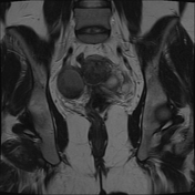
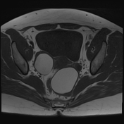
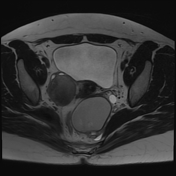
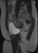
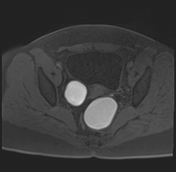
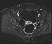
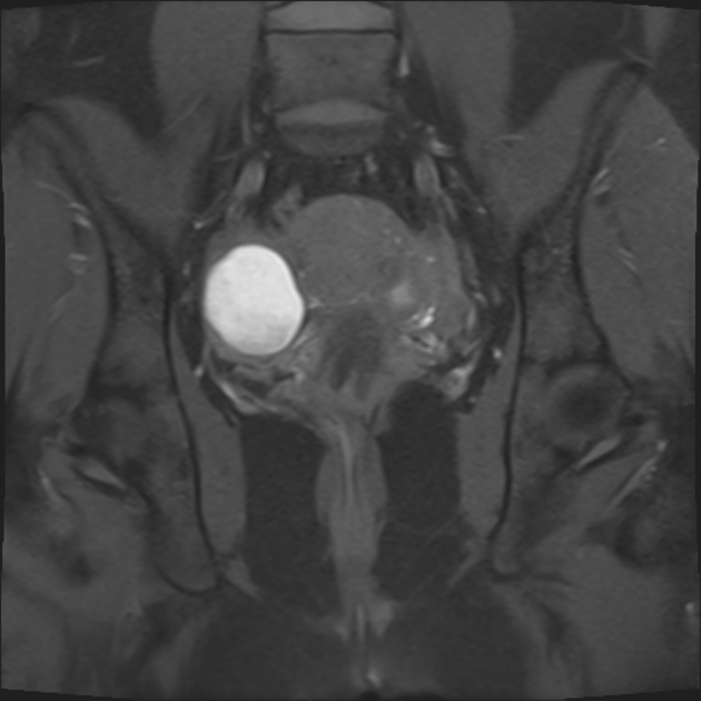
Both ovaries are enlarged containing multiple multiseptated cystic structures, one on the right side measuring 4.4 x 3.6 x 4.4 cm and two adjacent to each other on the left side measuring collectively 7 x 5 x 6.2 cm.
They demonstrate high signal intensity on T1WI fat sat and low signal intensity on T2WI (shading sign) along with T2WI hypointense dots ( T2 dark spot sign) with no evidence of diffusion restriction or suspicious enhancement.
The left ovary is more directly located to the midline causing mass-effect on the sigmoid shifting it to the right side.
Case Discussion
The patient went through laparoscopic surgery which confirmed the diagnosis. Radiological features help to establish the diagnosis include shading sign and T2 dark spot sign.


 Unable to process the form. Check for errors and try again.