Presentation
Painless swelling of the lateral aspect of the right elbow.
Patient Data
![](https://prod-images-static.radiopaedia.org/images/52848094/77ec5e5e97542c03991c64d2423ce4_thumb.jpeg)
![This study is a stack](/packs/stack-YQKLCKBI.gif)
![](https://prod-images-static.radiopaedia.org/images/52848179/f45e408782a54e39703e7653d128fd_thumb.jpeg)
![This study is a stack](/packs/stack-YQKLCKBI.gif)
![](https://prod-images-static.radiopaedia.org/images/52848206/c4813987165b63213d12fd6070bb28_thumb.jpeg)
![This study is a stack](/packs/stack-YQKLCKBI.gif)
![](https://prod-images-static.radiopaedia.org/images/52848131/041a747a2a9f4a246b68bb4ac52b82_thumb.jpeg)
![This study is a stack](/packs/stack-YQKLCKBI.gif)
![](https://prod-images-static.radiopaedia.org/images/52848228/3a9659e967a27bd43f1328534eaded_thumb.jpeg)
![This study is a stack](/packs/stack-YQKLCKBI.gif)
![](https://prod-images-static.radiopaedia.org/images/52848246/c4b9e56ed4fab9c5e9a6b4ff3db431_thumb.jpeg)
![This study is a stack](/packs/stack-YQKLCKBI.gif)
![](https://prod-images-static.radiopaedia.org/images/52848265/0fe046977cc80b13ff17c834a0362f_thumb.jpeg)
![This study is a stack](/packs/stack-YQKLCKBI.gif)
![](https://prod-images-static.radiopaedia.org/images/52848094/77ec5e5e97542c03991c64d2423ce4_big_gallery.jpeg)
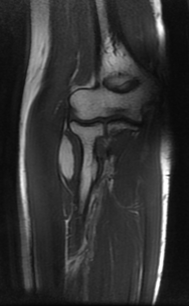
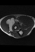
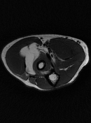
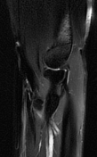
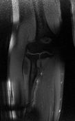
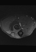
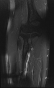
The MRI sequences demonstrate a well-defined lobulated deep soft tissue mass of intramuscular location within the supinator muscle with intermuscular extension displacing the brachioradialis and extensor carpi radialis longus muscles as well as the adjacent segment of the radial nerve. It displays a high signal on both T1WI and T2WI, attenuated on fat-saturated sequences. No enhancement seen on postcontrast sequences. No bony lesion or soft tissue infiltration. No joint effusion is seen.
Case Discussion
MRI features are most consistent of a soft tissues lipoma