
Patient Data
Age: 8 years
Gender: Female

Download
Info
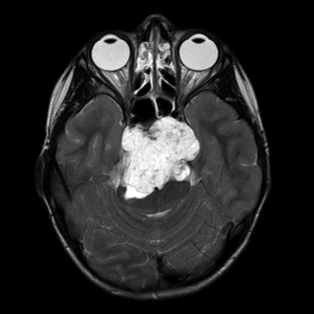
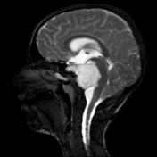
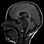
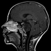
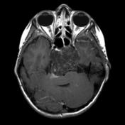
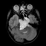
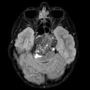
Large retroclival prepontine mass infiltrating the posterior aspect of the clivus and compressing the brain stem posteriorly. It is hypointense in T1, markedly hyperintense in T2 and shows minimal enhancement post contrast.
Case Discussion
This lesion was resected and confirmed to be a chordoma.


 Unable to process the form. Check for errors and try again.