
Presentation
Right neck mass.
Patient Data
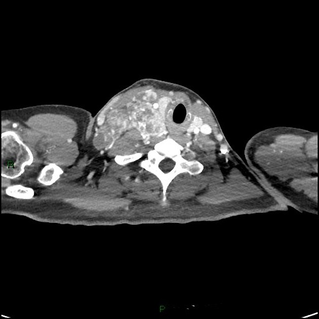
Several small nodules in the right lobe of the thyroid, all less than 1 cm in size.
Large volume right-sided and superior mediastinal lymphadenopathy. The nodes enhance avidly with extensive cystic areas within.
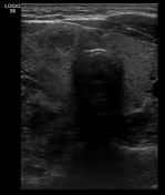
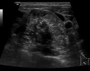
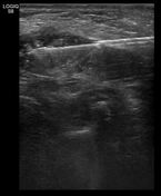

1 cm low echogenicity nodule with an instinct outline in the right lobe of the thyroid (U5).
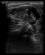
Large solid-cystic pathological neck nodes.
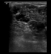
A core biopsy of a node was undertaken.
Case Discussion
The CT and ultrasound appearances of these metastatic nodes are different in appearance from the typical of metastatic nodes in the neck from the likes of a nasopharyngeal malignancy, lung or gastrointestinal tract malignancy. These are typical of from a papillary thyroid primary.
Note the tiny tumor in relation to the large volume metastatic lymph node disease. I have observed this several times with papillary carcinoma of the thyroid.
This patient falls into the most common group to suffer from this disease a female in the 3rd or 4th decade of life.


 Unable to process the form. Check for errors and try again.