
Presentation
Admitted in the emergency department for a three-week long headache and more recent dizziness, diplopia and hypoalgesia of the right trigeminal nerve.
Patient Data


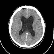
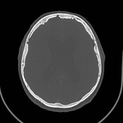

Bilateral contrast enhancing masses in both cerebellopontine angles that remodellate IACs with smooth-corticated edges, consistent with bilateral VIII craneal pair schwannomas.
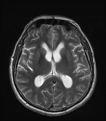
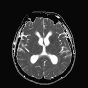
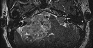
The right mass shows heterogeneous enhancement, causes infratentorial contralateral displacement and occupies ipsilateral basal cisterns with obstructive hydrocephalus signs, such as loss of the convexity sulci and dilatation of third and lateral ventricles.

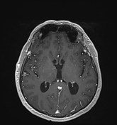

Both masses are well circumscribed, displace adjacent structures without direct invasion and extend into both IACs with meatal component.
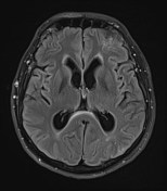
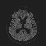
Right schwannoma shows intralesional hemorragic foci and cystic degenerative areas. It associates chronic ischemic areas in the ipsilateral cerebellar hemisphere, probably due to the compression made by the mass. It also causes obstructive hydrocephalus with the features described before, plus an Evans index of 0,43 and transependymal edema signs around the lateral ventricles.
A T2-FLAIR hyperintense area in the right frontal white matter can also be noted, extending from periventricular to subcortical region with no gadolinium enhancement. In this context it may be consistent with a glial microhamartoma.
Case Discussion
Bilateral vestibular schwannomas that cause obstructive hydrocephalus in a patient with a genetic diagnosis of neurofibromatosis type 2.


 Unable to process the form. Check for errors and try again.