
Presentation
Derranged LFTs and distended abdomen. No prior medical history.
Patient Data
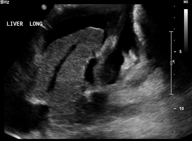
The liver is small, has coarse echotexture, and irregular contours consistent with cirrhosis. Small calcified left hepatic lobe granulomas, no suspicious liver lesions.
The portal vein is patent and has anterograde flow. Hepatic veins are patent but show decrease in phasicity and truncation of the a wave. Recanalization of the paraumbilical vein.
Spleen is not enlarged.
Large amount of ascites and left pleural effusion.
Case Discussion
Sonographic features are consistent with cirrhosis: small volume, irregular liver contours, and coarse echotexture. On Doppler, hepatic veins show decreased phasicity commonly seen in cirrhosis.
Recanalization of the paraumbilical vein and ascites support portal hypertension.


 Unable to process the form. Check for errors and try again.