
Presentation
Dry Cough, mixed obstructive/restrictive pattern on PFTs. Ex smoker
Patient Data

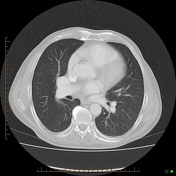
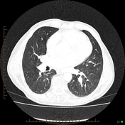

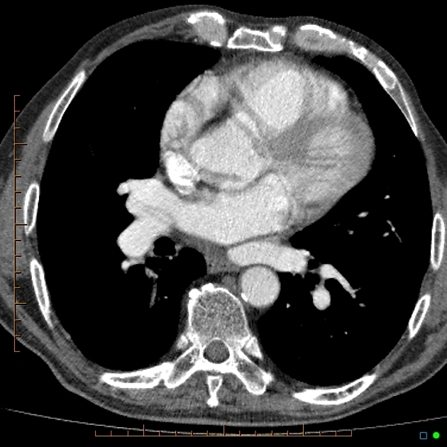
There are mild COAD changes in the lungs. No confluent airspace consolidation or any focal suspicious lesion in this smoker.
If you have already made the diagnosis, excellent!! Otherwise please scroll down to thin soft tissue windows.
I thought history was important as the findings are very unusual for patient's age and presentation.
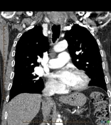
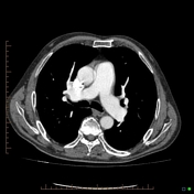
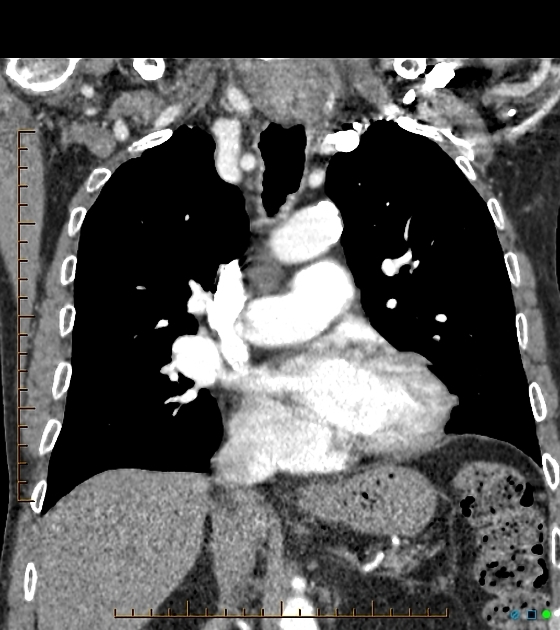
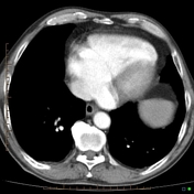
Partial anomalous pulmonary venous return of entire left lung. Please see below for the annotated images.
Also, bovine arch, a commonly seen variant.
Pulmonary veins from the left lung carrying oxygenated blood (therefore marked in red) drain into the right atrium via coronary sinus, resulting in a left to right shunt..
Case Discussion
This 70-year-old man has been pumping half of his oxygenated blood back to his right heart for 70 years now. Still, he has remained relatively asymptomatic despite being a smoker—one of the very rare cases of late diagnosis of PAPVR despite such a large shunt.


 Unable to process the form. Check for errors and try again.