
Presentation
History of swelling of the third digit of the right hand for 6 years.
Patient Data
Note: This case has been tagged as "legacy" as it no longer meets image preparation and/or other case publication guidelines.
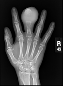
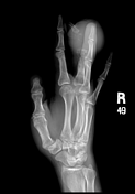


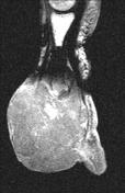
A destructive soft tissue mass lesion is noted epicentered on the middle phalanx of the right hand, which shows near total destruction. Its remnants show expansion and marked osteolytic changes. This is also involving the distal half of the proximal phalanx, showing mild expansion, osteolysis with prominent trabeculae.

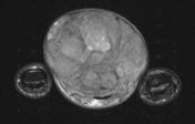
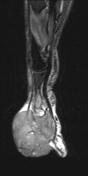

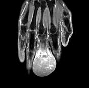
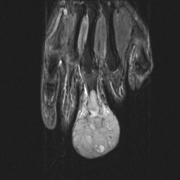
Soft tissue mass lesion is noted replacing the middle phalanx of the middle finger of the right hand with intense postcontrast enhancement. It is also involving the medullary cavity of the proximal phalanx.
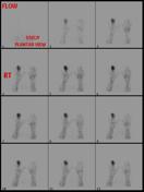
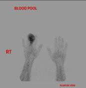
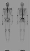
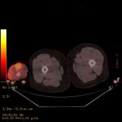

Increased uptake of the middle finger osteolytic destructive soft tissue mass lesion.
Case Discussion
Pathologically proved Ewing sarcoma.
Ewing's sarcoma of the finger is very rare and, pain & swelling of the affected finger are the most frequent presenting features.


 Unable to process the form. Check for errors and try again.