
Presentation
Fever diarrhea and malaise since two weeks ago, she denies abdominal pain or anorexia
Patient Data
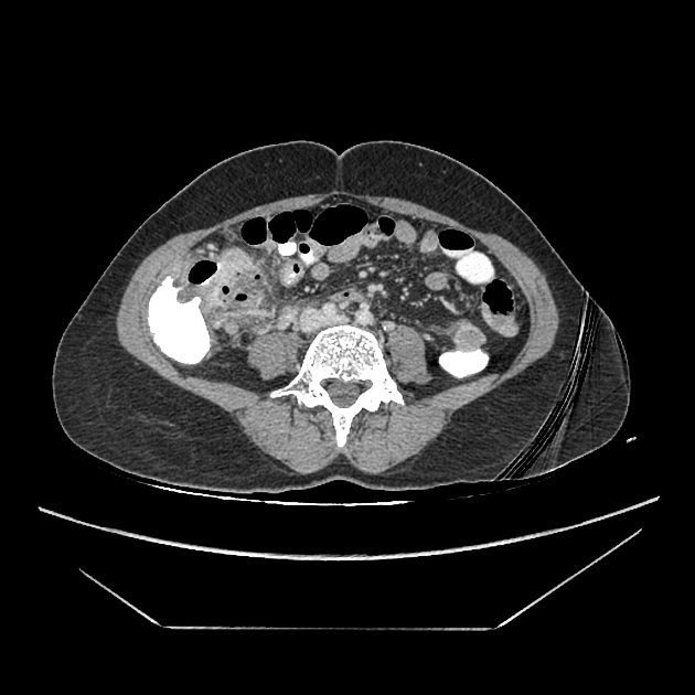
An abscess cavity containing air bubbles is seen posterior to the terminal ileum and reactive cecal wall thickening and small reactive lymph nodes. A short tubular structure representing inflamed proximal appendix is seen, arising from cecal tip merging with the abscess. Small elongated soft tissue posterosuperior to the abscess may be inflamed appendicular tip.
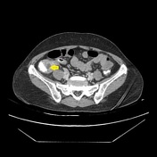
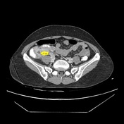
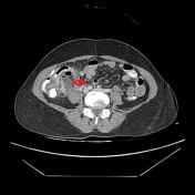

The red arrow points to the appendicular abscess, and yellow arrows show visible parts of the proximal inflamed appendix.
Case Discussion
The patient underwent percutaneous drainage of the abscess and received intravenous antibiotics. Colonoscopy was normal, and appendectomy was performed 8 weeks after the CT scan. The pathology report was in favor of appendicitis, and no mass lesion was detected.


 Unable to process the form. Check for errors and try again.