
Presentation
IVDU presenting with left-sided chest pain.
Patient Data
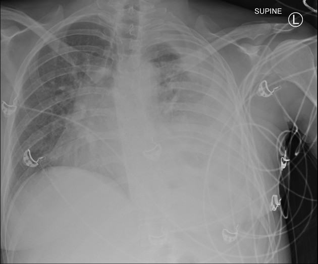
Almost complete whiteout of the left hemithorax. The mild shift of the mediastinal structures towards the right is equivocal if positional related. Features are supportive of a large pleural fluid collection. The lung and pleural space on the right appear clear. No evidence of pneumothorax.


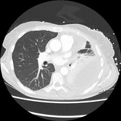
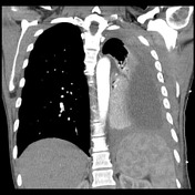
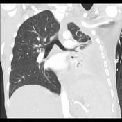
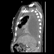
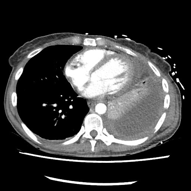
Moderate volume left-sided low density pleural fluid with diffusely thickened pleura and forming obtuse angles with the adjacent lung. A split pleural sign is noted. No appreciable gas within the collection. Findings are suggestive of pleural empyema.
Associated segmental compressive atelectasis and, perhaps, a small component of consolidation, most pronounced within the left lower lobe.
Mild right basal atelectasis. The right lung and pleural space are clear.
Small volume pericardial effusion. Otherwise normal appearance of the mediastinal soft tissue and vascular structures. No appreciable mediastinal, hilar or axillary lymphadenopathy.
No suspicious osseous lesion.
Case Discussion
Features are those of a thoracic/pleural empyema. This case illustrates the split pleural sign, where fibrin coating both the parietal and visceral surfaces of the pleura allow them to be visualized as linear regions of enhancement that come together at the margins of the collection.


 Unable to process the form. Check for errors and try again.