
Presentation
Pain in right hypochondrium.
Patient Data
Age: 70 years
Gender: Female
From the case:
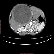
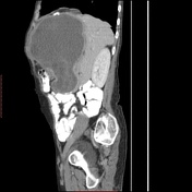
Hepatic hydatid cyst


Download
Info
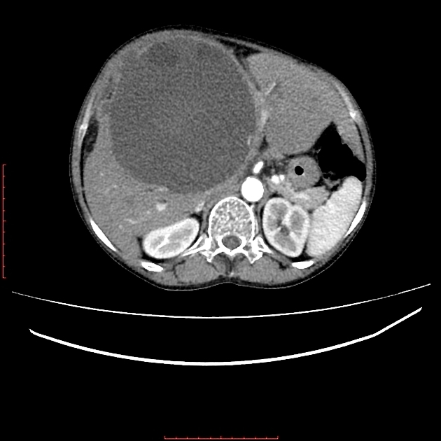
A large well-defined cystic lesion in the right hepatic lobe showing multiple small daughter cysts and wall calcifications. Note the smaller exophytic component of this cyst along the anteroinferior aspect abutting the adjacent small and large bowel loops. Loculated fluid collection is also seen in the right sub-diaphragmatic space.
Case Discussion
Hydatid cyst occurs secondary to the infection by the Echinococcus species. It is most commonly seen involving the liver followed by lungs and spleen.
Due to the huge size of the cyst, this patient underwent surgical excision and is doing good on follow-up.


 Unable to process the form. Check for errors and try again.