
Presentation
Abdominal pain.
Patient Data


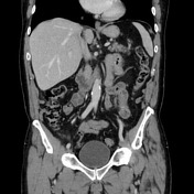
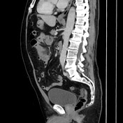
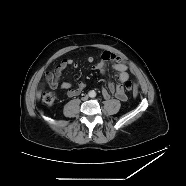
Very large, inflamed jejunal diverticulum in the left mid abdomen. Notice communication with the bowel lumen. Extensive fat stranding. Small oval enhancing collection along the medial aspect could be a small abscess or reactive inflammation of a separate diverticulum. Several other small, non inflamed diverticula along the mesenteric border of the jejunum.
Right inguinal hernia with flat, fluid, and soft tissue thickening.
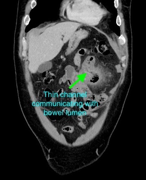
Annotated image showing communication of the diverticulum with the bowel lumen.
Case Discussion
The important thing when reading this case is to not call this an abscess. It is clearly communicating with the small bowel lumen, and upon closer inspection there are several other smaller non-inflamed jejunal diverticula along the mesenteric border. Thus, it is important to be familiar with the appearance of small bowel diverticulitis, which often presents with much larger and more impressively inflamed diverticula in the sigmoid colon. The diagnosis can be supported and confidently made when observing the presence of other non-inflamed small bowel diverticula.


 Unable to process the form. Check for errors and try again.