
Presentation
MVA, patient in collar. CT C spine as part of trauma workup.
Patient Data
Age: 30 years
Gender: Male
From the case:
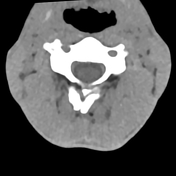
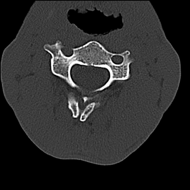
External laryngocoele and pharyngocoele


Download
Info
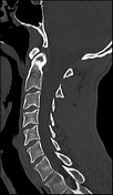
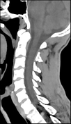
Vertebral body alignment and vertebral body height is maintained. No cervical spine fracture.
Small incidental right-sided external laryngocoele. It appears to extend superiorly around the hyoid bone and communicating with the right vallecula representing a pharyngocoele. No laryngeal mucosal lesion identified at the opening of the ventricle.
Large CSF density space anterior to the left temporal pole likely represents an arachnoid cyst.
Case Discussion
This is a peculiar case of a combined laryngocoele and pharyngocoele.


 Unable to process the form. Check for errors and try again.