
Patient Data
Age: 16 years
Gender: Male
From the case:
Myositis ossificans - arm
Download
Info
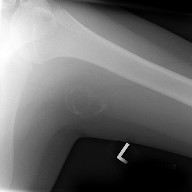
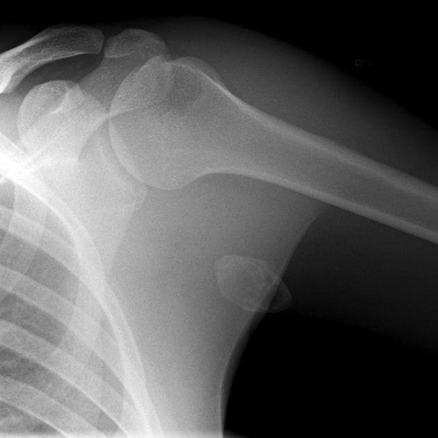
Well circumscribed peripherally calcified lesion, separate from the underlying humerus which appears normal.
From the case:
Myositis ossificans - arm
Download
Info

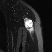
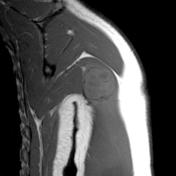
MRI demonstrates heterogeneous signal generally isointense to that of muscle on T1, and heterogeneous high signal on T2 weighted images.
From the case:
Myositis ossificans - arm
Download
Info
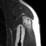
Increased calcification and decreased size (expected evolution) one year later.
Case Discussion
Findings are in keeping with myositis ossificans.


 Unable to process the form. Check for errors and try again.