
Presentation
Known renal cell carcinoma on treatment. Recent confusion.
Patient Data
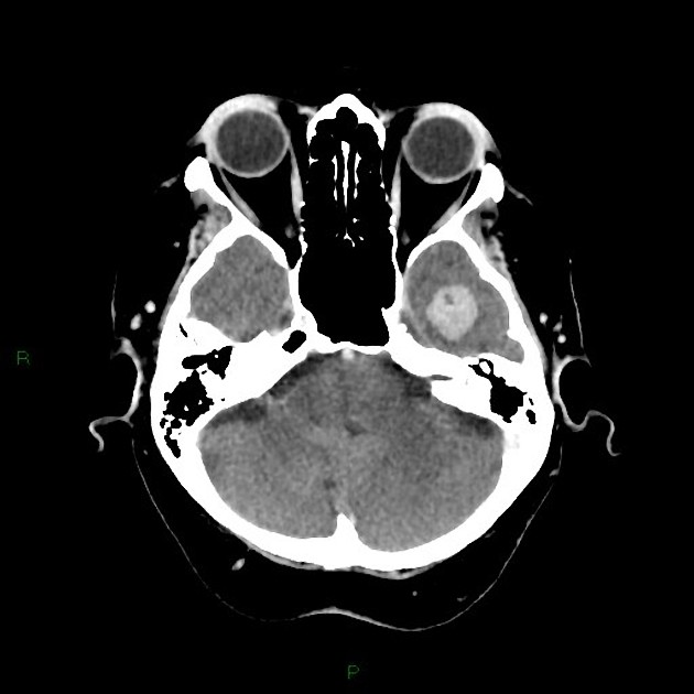
Solitary avidly enhancing left temporal lobe metastasis. No perilesional edema.
The remainder of the intracranial appearances are normal.
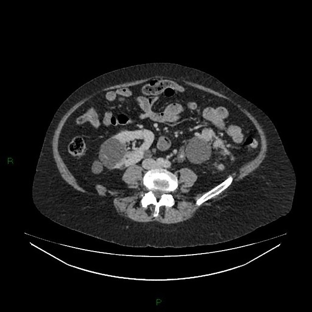
7cm mass with central necrosis in the mid and lower pole of the left kidney. The mass extends into the lower pole branch of the left renal vein and extends beyond the capsule to abut Gerota's fascia.
Simple cysts in both kidneys.
No solid organ, adrenal or peritoneal metastases.
No infradiaphragmatic lymphadenopathy. No bone lesion.
Adnexal cyst in the right side of the pelvis.
Case Discussion
Renal cell carcinoma metastases as in this case are typically hypervascular as with the primary tumor.
The radiological stage for this case T3a,N0,M1 (brain).
The T3a stage relates to both its extension into segmental veins and the perirenal tissues.


 Unable to process the form. Check for errors and try again.