
Presentation
Progressive dyspnoe, fatigue low SpO2. Minimally increased inflammatory markers. Pneumonia?
Patient Data
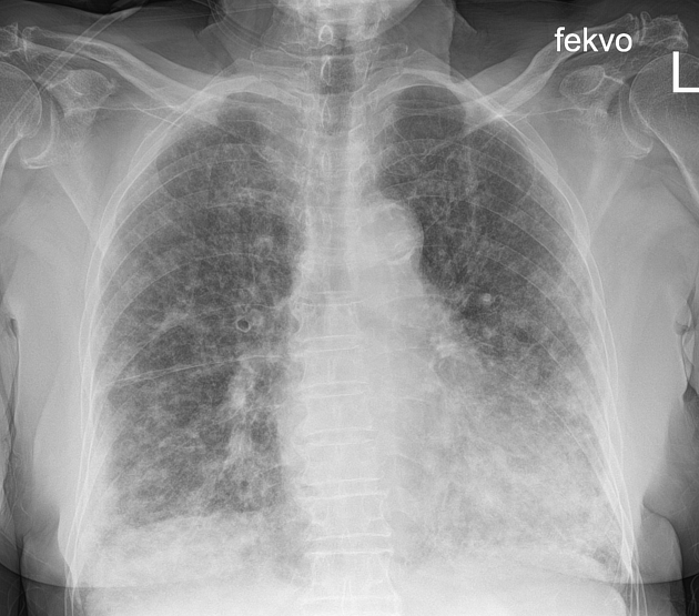
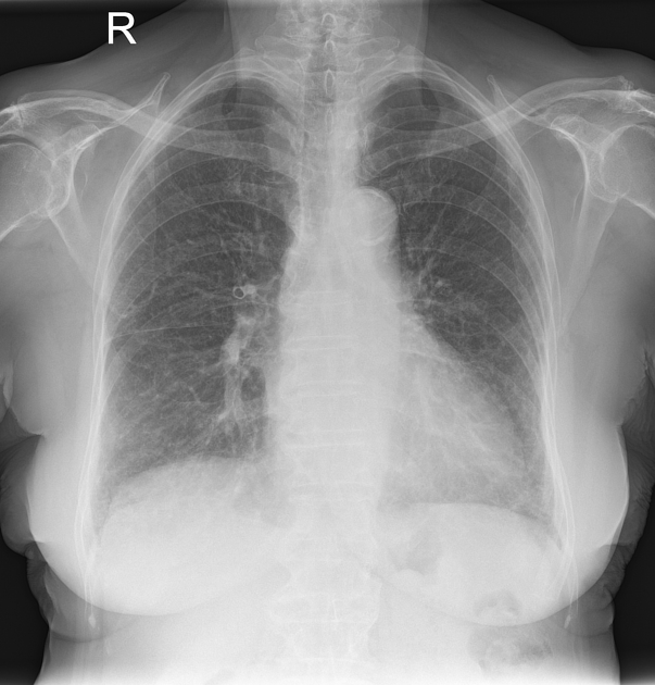
Bilateral coarse, patchy reticulation with a marked basal and peripheral predominance. Novel when compared to prior radiograph. Atypical pneumonia or interstitial process suggested.
Antibiotic therapy was initiated but without clinical or radiographic improvement over the following days. Considering the ongoing medications of the patient and the lack of elevated inflammatory markers the possibility of amiodarone-induced fibrosis was raised, and a CT was requested 10 days later.


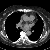
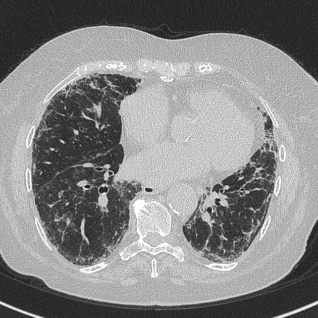
Bilateral, predominantly subpleural, bibasal reticulation with moderate traction bronchiectasis, and small ground glass opacities. Findings are suggestive of usual interstitial pneumonia.
The amiodarone therapy was subsequently discontinued and steroid medication was initiated, followed by gradual clinical improvement over the following weeks.
Note: the number of slices has been reduced.

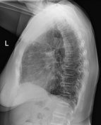
Marked improvement when compared to prior exams.
Case Discussion
Peripheral air-space opacities and subpleural fibrosis are typical consequences of amiodarone pulmonary toxicity, but the radiographic appearance alone is unfortunately far from pathognomonic. Both the progression of the disease and recovery following the discontinuation of the drug and initiation of non-specific anti-inflammatory therapy can be rapid.


 Unable to process the form. Check for errors and try again.