
Presentation
SOB on a background of COPD.
Patient Data
Age: 60 years
Gender: Male
From the case:
COPD - pulmonary bullae

Download
Info

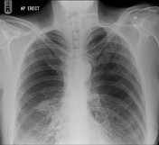
Large bullae to both superior and mid zones. Severe chronic emphysematous changes are noted at both lung fields. Increased air space opacification right mid and lower zones.
Case Discussion
It can sometimes be challenging to distinguish between bullae and a pneumothorax on a plain film. CT chest is often used in the emergency department to differentiate between pneumothorax and a large bullous before attempting a chest drain insertion.


 Unable to process the form. Check for errors and try again.