
Presentation
Abdominal pain.
Patient Data
Age: 65 years
Gender: Male
From the case:
Pulmonary, liver and spleen hydatid cysts


Download
Info
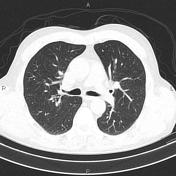
Two cystic lesions are seen at right and left lung bases measuring 75×63mm and 28×23mm, respectively. Additionally, mild focal bronchiectasis is present at left lower lingular segment.
Degenerative changes as osteophytosis are seen at the thoracic spine.
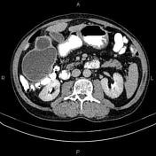
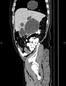
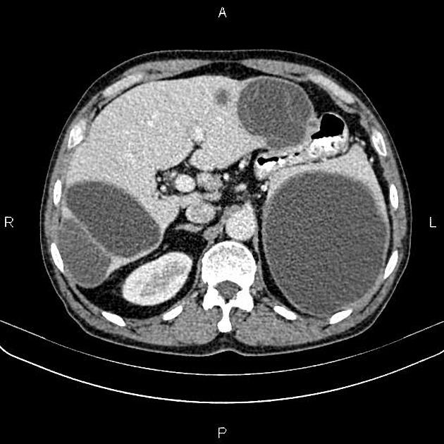
Multiple thick-walled cystic lesions with internal layer detachment are seen at liver. The largest one measured 100×80mm.
A 125×115mm thick walled cystic lesion is present at spleen. No calcification or enhancing solid component are seen within.
Several non-enhanced simple cortical cysts are seen at both kidneys, with maximum diameters of 35mm.
Case Discussion
Features on CT scan are compatible with pulmonary, hepatic & splenic hydatid cysts.


 Unable to process the form. Check for errors and try again.