
Presentation
Dialyzing left jugular CVC inserted elsewhere, sharp chest paint immediately after beginning of dialysis, approximately 1L dialyzing fluid likely paravasated. Prior CT at referring hospital showed no acute bleeding. Sinking Hgb levels. Active bleeding?
Patient Data
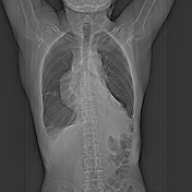
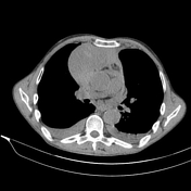

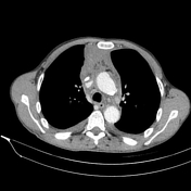
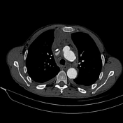
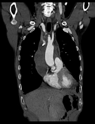


- Large, approximately 110 x 110 x 70 mm inhomogeneous anterior mediastinal collection, adjacent to the extravasated CVC tip locally increased density is suggestive of sentinel clot.
- Bilateral pleural effusion (increased density) and resultant atelectasis.
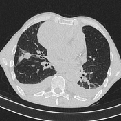
- Spiculated, known pulmonary mass on the right with satellite nodules in keeping with malignancy.
- Subtle pericardial effusion.
- Avidly enhancing small lesion in the 4A segment of the liver can be fast-filling hemangioma, but the chest angio CT exam is not suitable for complete characterization
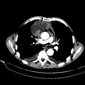
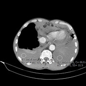

Key images demonstrating the sentinel clot and bilateral high-density pleural effusion.
Case Discussion
While the large mediastinal collection and the catheter-tip extravasation are self-evident, the subtle sentinel clot sign and the relatively high density of the bilateral pleural effusion are less so at first glance. Nonetheless these key features show that there is slow but active venous bleeding.
Active venous bleeding and resultant hemothorax was confirmed during subsequent operation.


 Unable to process the form. Check for errors and try again.