
Presentation
Occipital swelling.
Patient Data


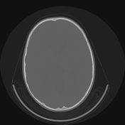
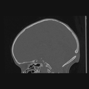
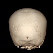

There is a well-marginated defect with a thin sclerotic border in the midline of the occipital bone measuring 8.5 x 3.3 mm. Superficial to the defect, a well-defined ovoid heterogeneous lesion with areas of fluid attenuation is seen in the scalp measuring 1.8 x 0.8 x 1.9 cm, which appears to communicate with the intracranial space.
Enlarged retrocerebellar CSF space with an intact cerebellar vermis. No communication with the fourth ventricle is seen. Findings are compatible with a mega cisternal magna.

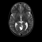
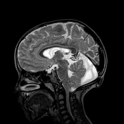
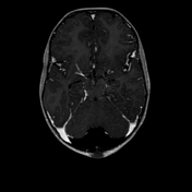
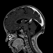
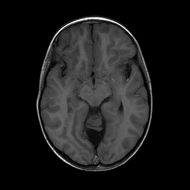
There is a small midline occipital scalp lesion measuring about 2 x 1.6 x 0.8 cm connected to the brain through a small midline occipital bone defect measuring about 8 x 4 mm. It appears heterogeneous and of mixed-signal intensity mainly in T2 images. No vessels, fat, or significant enhancement seen in the mentioned lesion.
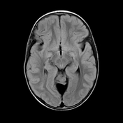
No associated intracranial abnormalities.
Case Discussion
Cephaloceles represent herniated intracranial content through a skull defect.
Atretic cephaloceles are involuted cephaloceles containing meninges and neural crests and represent about 50% of all cephaloceles.
The majority of reported cases occurred in the parietal region, although some reports suggest equal incidence on both sides of the lambdoid suture (parietal and occipital).
An increased incidence of intracranial anomalies has been reported with atretic cephalocele, and these include Dandy-Walker syndrome, Walker-Warburg syndrome, and holoprosencephaly, among others.
Our patient went on to have surgical excision of the cephalocele. Histopathology found meningeal tissue within.


 Unable to process the form. Check for errors and try again.