
Presentation
Abdominal discomfort.
Patient Data


Partially images calcifications in the lung base.
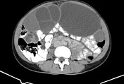
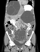
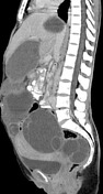
Large cyst with daughter cysts right hepatic lobe. Atrophy of much of the left hepatic lobe, with adjacent cyst containing subtle amounts of fat suggesting communication with the biliary tree.
Numerous cysts of various sizes throughout the peritoneum and pelvis, with variable density, daughter cysts, and few small peripheral calcifications. Mild right hydrouteronephrosis due to compression by the larger pelvic cyst.
Case Discussion
Very large burden of liver and peritoneal hydatid disease with various stages, although many are active and characterized by the presence of daughter cysts. This presumably began as a liver cyst (most common site to become infected) which eventually ruptured and seeded the peritoneal cavity.


 Unable to process the form. Check for errors and try again.