Presentation
Acute low back pain.
Patient Data
![](https://prod-images-static.radiopaedia.org/images/53706325/664fae0ba2b39580914fcd11de5ceea7f5084940f071d92fc1e71994efacb335_thumb.jpeg)
![This study is a stack](/packs/stack-YQKLCKBI.gif)
![](https://prod-images-static.radiopaedia.org/images/53706635/2cd47396e9cdfbf4159512513e63e26388760de19bf4ed9c5a9a5ccb43b8080c_thumb.jpeg)
![This study is a stack](/packs/stack-YQKLCKBI.gif)
![](https://prod-images-static.radiopaedia.org/images/53706726/2dcc83fd69e4316e70895e0bc93c715040c3b04e1f5385c3465953901c65de32_thumb.jpeg)
![This study is a stack](/packs/stack-YQKLCKBI.gif)
![](https://prod-images-static.radiopaedia.org/images/53706825/066710f251b752adbdc3e0d1aca146deb3c81b6b85e122062e1bf8886677a800_thumb.jpeg)
![This study is a stack](/packs/stack-YQKLCKBI.gif)
![](https://prod-images-static.radiopaedia.org/images/53706325/664fae0ba2b39580914fcd11de5ceea7f5084940f071d92fc1e71994efacb335_big_gallery.jpeg)
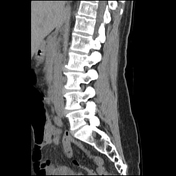
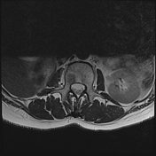
Lumbar scoliosis. Lucency within the L2 superior endplate with cortical erosion. No other focal lesion. Right nephrectomy.
![](https://prod-images-static.radiopaedia.org/images/53706868/3e40f44f62cdeaccafa0aec0267ff9a9ad14dedf843a01f280440d1460c417ae_thumb.jpeg)
![This study is a stack](/packs/stack-YQKLCKBI.gif)
![](https://prod-images-static.radiopaedia.org/images/53706505/3b5691d9dfb2f7451ae2ef2910d0b09fc7257dcbd9d6a6718c239c5e97c94253_thumb.jpeg)
![This study is a stack](/packs/stack-YQKLCKBI.gif)
![](https://prod-images-static.radiopaedia.org/images/53706526/65e15555e52fc656819778d5d60c9b02b80ce3d3cc0a2dc45e70844d3e202e44_thumb.jpeg)
![This study is a stack](/packs/stack-YQKLCKBI.gif)
![](https://prod-images-static.radiopaedia.org/images/53706763/73d524b4240c1efa4d04d53f5d30004ba12004e0e32c7e9262563731444158b0_thumb.jpeg)
![This study is a stack](/packs/stack-YQKLCKBI.gif)
![](https://prod-images-static.radiopaedia.org/images/53706580/cbbede5cc29de470c5148b7b5aa1a4dcd8805ed1a3f39f69f7b9b5af9a513d50_thumb.jpeg)
![This study is a stack](/packs/stack-YQKLCKBI.gif)
![](https://prod-images-static.radiopaedia.org/images/53706554/98327f3a6d66f568041f6da2d8c93d9813c7f0c652dc62f3b2b5d29d34360d3a_thumb.jpeg)
![This study is a stack](/packs/stack-YQKLCKBI.gif)
![](https://prod-images-static.radiopaedia.org/images/53706444/87ffc54b3f5100d34136aa5ef6cb4943b481d9558fe8dd954a0646998f9d6734_thumb.jpeg)
![This study is a stack](/packs/stack-YQKLCKBI.gif)
![](https://prod-images-static.radiopaedia.org/images/53706783/11b2eb82ef2f4fc6469f7e92a1c6a9f81744a2f1f8cc2b5e2deb31bb9741b2c5_thumb.jpeg)
![This study is a stack](/packs/stack-YQKLCKBI.gif)
![](https://prod-images-static.radiopaedia.org/images/53706686/c15a8f24f18b2397941b4e94a841d8497ea651bd7b3c1ea9a6ab3ad88e970415_thumb.jpeg)
![This study is a stack](/packs/stack-YQKLCKBI.gif)
![](https://prod-images-static.radiopaedia.org/images/53706868/3e40f44f62cdeaccafa0aec0267ff9a9ad14dedf843a01f280440d1460c417ae_big_gallery.jpeg)
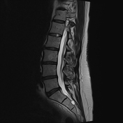
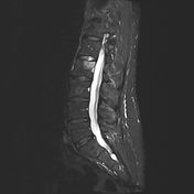
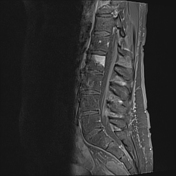
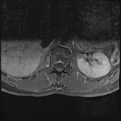
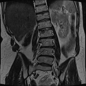
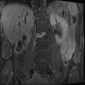
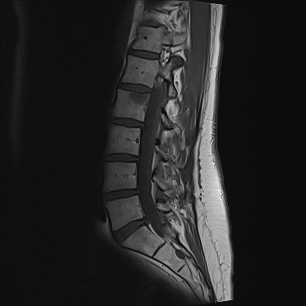
At the right aspect of the L2 vertebral body just inferior to the endplate is a region of bone marrow edema centered on a disrupted and slightly depressed L2 superior endplates with edema extending into the right pedicle. Postcontrast enhancement is relatively diffuse in this region with normal signal intervertebral disc extending inferiorly in keeping with a Schmorl's node. Mild periosteal and paravertebral soft-tissue edema.
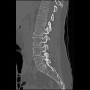
Mild L1/2 disc edema. No further focal osseous lesion is identified. Enhancement of the right L2 nerve root.
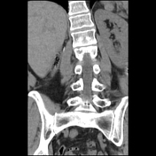
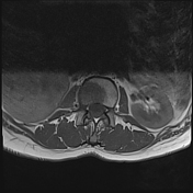
Previous right nephrectomy. Paraspinal soft tissues demonstrate no gross abnormality.
Case Discussion
Bone marrow edema and enhancement centered inferior to the right aspect of the L2 superior endplate is favored to be an acute Schmorl's node with reactive edema. The main differential is a vertebral metastasis but the contiguity with the intervertebral disc and similar signal heavily favor an acute Schmorl's node despite a previous right nephrectomy for renal cell carcinoma.