
Presentation
Right knee pain 1 year. Mild worsening of the pain associated with decreased range of motion over the last 6 months. No history of trauma, or fever.
Patient Data

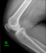
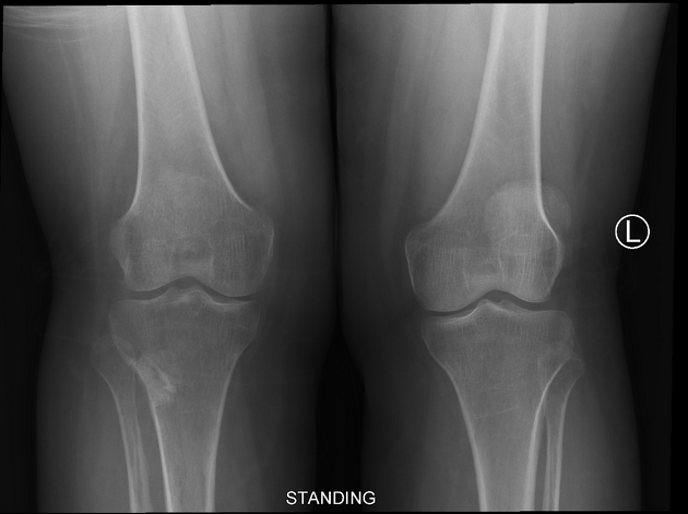
A focal radiolucent lesion with a thin peripheral sclerotic rim and narrow zone of transition is seen along the posterolateral aspect of the proximal metaphysis of the right tibia. Focal cortical thinning is seen along the lateral aspect of the proximal tibia; however, no periosteal reaction, fracture, or associated soft tissue abnormality is seen. These radiographic features are suggestive of a non-aggressive osseous lesion, like bone cyst.


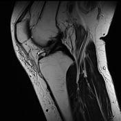
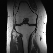
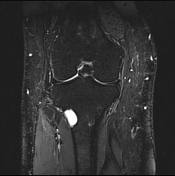
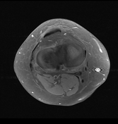
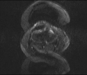
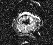
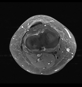
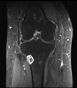
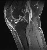

Findings: A well-defined eccentric unilocular cystic lesion having lobulated sclerotic margins is seen along the posterolateral aspect of the proximal metaphysis of the right tibia. Minimal cortical thinning is noted at the site of the lesion. No abnormal signal is seen in the adjacent soft tissues. No additional bony lesions are seen. The lesion shows peripheral enhancement on the post contrast study. No abnormal joint effusion is seen. Focal cartilaginous loss with subchondral cyst formation supportive of focal chondromalacia patella is noted. Mild increased signal is noted along the anterior aspect of the patellar ligament, which is suggestive of mild prepatellar bursitis.
Impression: Benign looking eccentric proximal tibial metaphyseal cystic lesion suggestive of intraosseous ganglion cyst. Differential diagnosis include atypical non-ossifying fibroma.
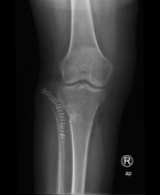
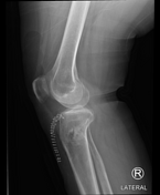

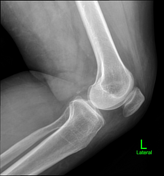
Evidence of curettage and bone grafting is noted at the site of radiolucent lesion in the proximal right tibia. Postsurgical changes are seen in the adjacent soft tissues.

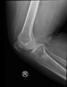
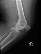
Interval healing is noted at the site of previous curettage and bone grafting of the right proximal tibial radiolucent lesion.
Case Discussion
Procedure: Tibial cyst curettage and bone grafting.
Histopathological analysis of tibial cyst curettage: Histological features of benign bone cyst consistent with an intraosseous ganglion cyst.


 Unable to process the form. Check for errors and try again.