
Chondromalacia patellae refers to softening and degeneration of the articular hyaline cartilage of the patella that articulates with the trochlear groove of the femur and is a frequent cause of anterior knee pain.
On this page:
Epidemiology
Tends to occur in young adults. There is a recognized female predilection.
Associations
Chondromalacia patellae can either occur in isolation or secondary to other conditions, including 1-4:
direct trauma
chronic patellar instability/subluxation
Clinical presentation
Patients with chondromalacia patellae usually present with anterior knee pain on walking up or down stairs. Additionally, there may be knee pain when kneeling, squatting, or after sitting for long periods of time. Knee stiffness, crepitus and effusions may also be present. In some cases, a history of patellar dislocation may be present 4. Some patients may present with quadriceps wasting or atrophy 10.
Radiographic features
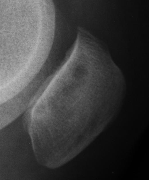
Plain radiograph
Plain radiographs of the knee cannot assess for chondral changes directly and can only demonstrate features of osteoarthritis (OA) involving the patellofemoral joint in end-stage disease. A joint effusion may be visible. Lateral and skyline views are more helpful to assess for shallow excavation in the subchondral bone involving the patella.
CT
CT arthrography can be used to diagnose plicae and focal cartilage defects but can be insensitive to early chondral injury 3.
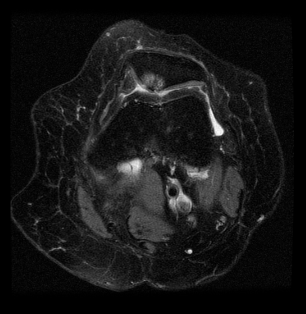
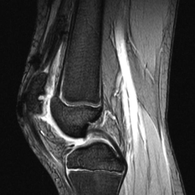
MRI
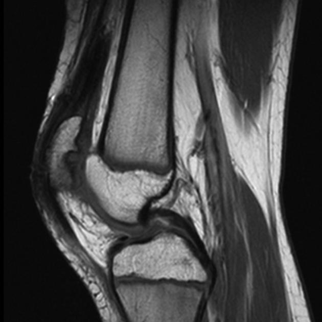
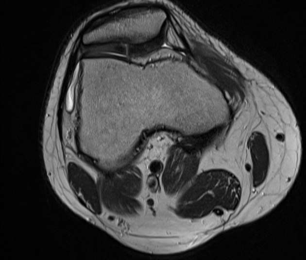
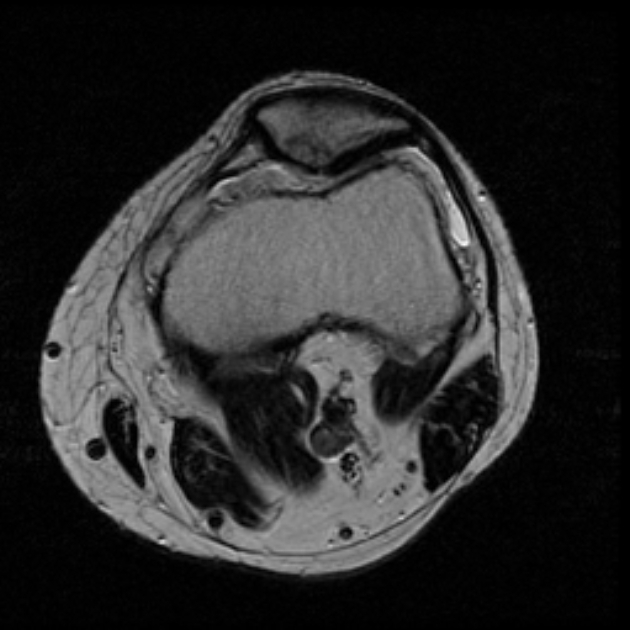
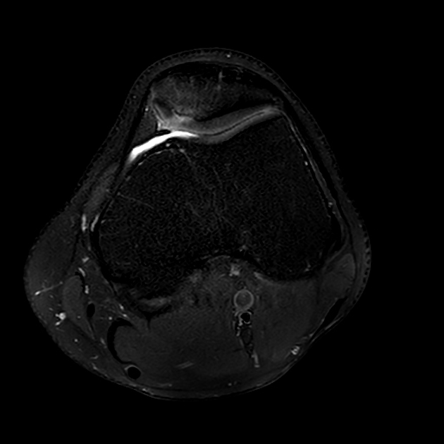
MRI is the modality of choice for assessing patellar cartilage.
-
T1
poor sequence for cartilage; surface irregularity and subtle signal change may be inapparent
areas of hypointensity may be seen in cartilage
hypointense subchondral reactive bone marrow edema pattern may be seen
secondary changes of osteoarthritis may be seen
-
T2/PD
best sequences for assessing cartilage
most patients with chondromalacia patellae have focally increased signal in the cartilage or focal contour defects in the cartilage surface
abnormal cartilage is usually high signal compared to normal cartilage
findings range from a subtle increase in signal to complete loss of cartilage
In the absence of an effusion, plicae may be difficult to identify 3.
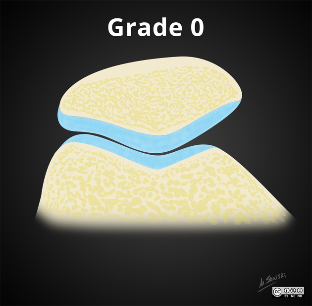
Grading
The grading system of chondromalacia patellae is based on T2/PD weighted MRI findings and arthroscopic correlation:
modified Outerbridge grading (most widely used)
Treatment and prognosis
Initial management is non-operative with a reduction of strenuous activities, NSAIDs, and exercises to stretch and strengthen the quadriceps muscle (especially vastus medialis) 4.
A variety of operative options exists including 4:
arthroscopic debridement and lavage: diagnostic but only offers short term symptomatic relief
articular resurfacing
surgical correction for instability
patellectomy
Differential diagnosis
General imaging differential considerations include:
dorsal defect of the patella: on the superolateral corner of the patella
-
bone marrow edema at the inferior pole as a result of


 Unable to process the form. Check for errors and try again.