
Presentation
Abdominopelvic mass, which is progressively increasing in size over the last two years with a positive past history of uterine fibroid. No urinary or bowel symptoms.
Patient Data

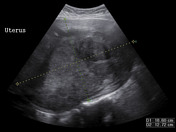
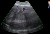
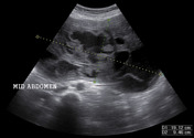
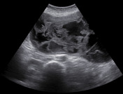
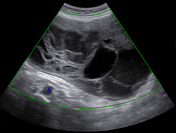
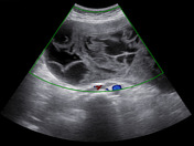
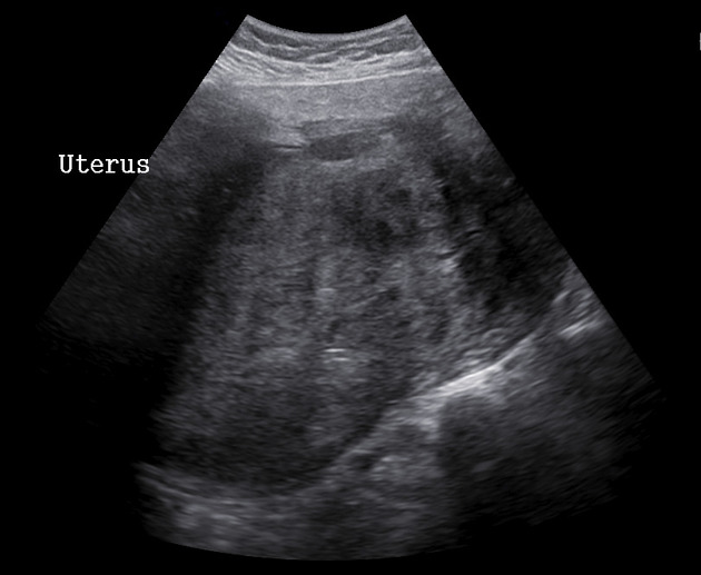
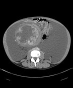
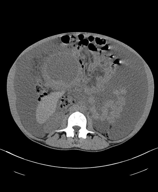
A large heterogeneous solid uterine mass measuring about 18.6 x 12.7 cm is seen in the pelvis. Another large mass containing mixed cystic and solid components measuring 19 x 9 cm is seen in the central abdomen. Both these masses are connected to each other. No internal vascularity is seen in these masses on color Doppler ultrasound examination.
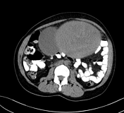

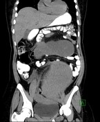
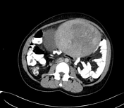
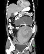

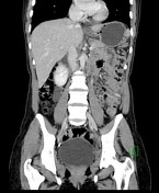
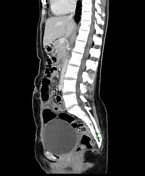
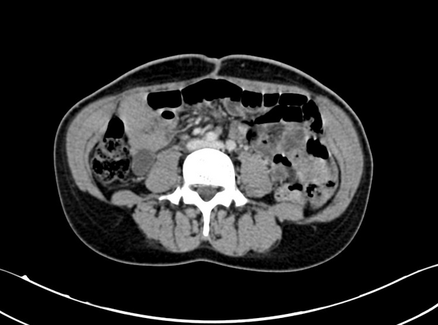
Huge abdominopelvic mass arising from the uterus. It appears bilobed in nature with its inferior component having mixed solid and cystic components (predominantly solid) whereas its superior component is predominantly cystic in nature and has a few thin enhancing septae. Mild ascites and a tiny cyst in the right hepatic lobe. No evidence of loco-regional or distant metastasis is seen.
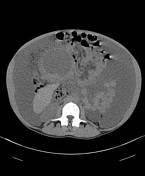


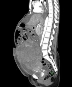


Large heterogeneous mixed solid and cystic uterine mass showing mild interval increase in its size when compared with the previous scan. Marked interval increase is seen in the ascites. Interval development of moderate right-sided pleural effusion and small umbilical and right inguinal hernias containing ascitic fluid.
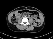
Status post hysterectomy and bilateral oophorectomy. No evidence of recurrence or metastases is seen. Stable tiny hepatic cyst.
Case Discussion
The patient was lost follow up after the initial work-up. Two years later, she presented with worsening abdominal distension, anorexia and weight loss. After repeat work-up, she underwent surgery.
Procedure: Total abdominal hysterectomy (TAH), bilateral salpingo-oophorectomy (BSO), omentectomy, bilateral parametrium removal and umbilical hernia repair.
TAH & BSO: Leiomyosarcoma. Size: 16 cm. Coagulative necrosis present. Mitotic rate= 7/10 HPF. Moderate nuclear atypia with focal severe nuclear atypia. Endometrium: Proliferative endometrium. Cervix: Mild chronic nonspecific inflammation. Fallopian tubes: Para tubal cyst with no other significant changes. Left ovary: Benign follicular cyst with no other significant pathology. Right ovary: No significant pathology.
Peritoneum & omentum: Chronic inflammation with hemosiderin deposition and reactive mesothelial hyperplasia. No evidence of malignancy seen.
Ascitic fluid analysis: Negative for malignant cells.


 Unable to process the form. Check for errors and try again.