
Presentation
Abdominal pain.
Patient Data
Age: 55 years
Gender: Male
From the case:
Pancreatic adenocarcinoma


Download
Info
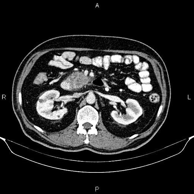
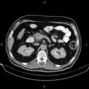
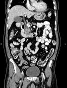
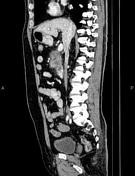
A 35 x 30 mm heterogeneously enhancing mass in the pancreatic head, appearing to infiltrate the adjacent duodenal wall, with mild surrounding fat stranding. There are several small lymph regional nodes. Parenchymal atrophic changes and main pancreatic duct dilatation distal to the mass.
A few sub-centimeter simple renal cortical cysts.
Case Discussion
Pancreatic head mass with small regional lymph nodes and suspected invasion to the adjacent duodenum. Pathology proven pancreatic adenocarcinoma.


 Unable to process the form. Check for errors and try again.