
Presentation
Painless neck progressive swelling.
Patient Data
Age: 45 years
Gender: Male
From the case:
Liposarcoma


Download
Info
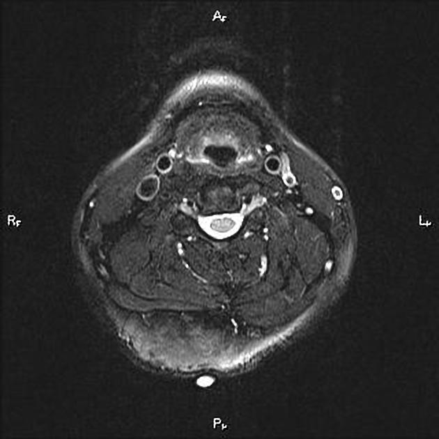
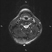
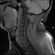
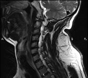
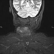
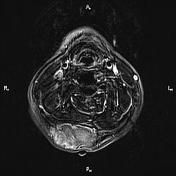
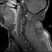
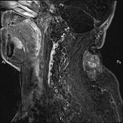
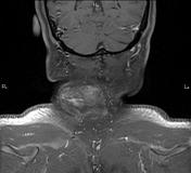
A 44×56×30mm subcutaneous mass with lobulated margin and several internal septations are seen at right posterior aspect of the lower neck.
The mass shows fat signal intensity on all pulse sequences.
There is no sign of local invasion to adjacent muscular structures.
After contrast media administration, the mass shows mild heterogeneous enhancement.
Case Discussion
Path proven cervical liposarcoma, which is malignant tumor of fatty tissue and is the malignant counterpart to a benign lipoma. Liposarcoma is the second most common type of soft-tissue sarcoma.


 Unable to process the form. Check for errors and try again.