
Presentation
Abdominopelvic mass feeling.
Patient Data
Focal course calcification is present at central portion of the pelvis.
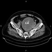


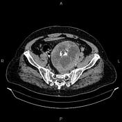
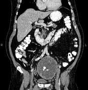
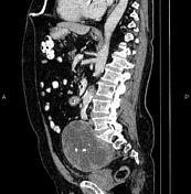

A 145×105×115mm mass with central coarse calcification and heterogeneous enhancement is present in the midline pelvis, anterior to the sacrum, and superoposterior to the urinary bladder. There is no sign of local invasion to adjacent pelvic structures, no bony sacral destruction, and no intraspinal extension.
A few subcentimeter simple cortical cysts are seen in kidneys.
The prostate gland is markedly enlarged.
Case Discussion
Pathologically proved pelvic ganglioneuroma.
Ganglioneuromas are usually asymptomatic and often discovered incidentally as they are slow-growing and generally endocrinologically inactive 1. They are typically seen as well-circumscribed encapsulated masses that are iso to hypoattenuating to muscle. The lesion may also demonstrate calcifications (~20%) which are generally fine and speckled but may be coarse 2.


 Unable to process the form. Check for errors and try again.