
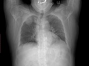
Presentation
Dyspnea
Patient Data
Age: 70 years
Gender: Male
From the case:
Pleural lipoma


Download
Info

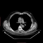
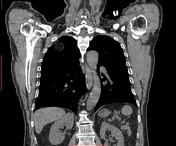
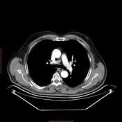
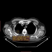
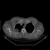
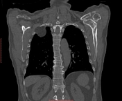
A well-circumscribed non-enhancing convex pleural-based lesion forming obtuse angles with the pleura is noted in the right hemithorax contiguous to the posterior segment of the right upper lung lobe and contacting the right 2nd and 3rd ribs posteriorly with no detected rib erosion. The mean attenuation value was -120 HU (denoting fat).
Case Discussion
As usual, this pleural lipoma was incidentally discovered during CT chest.
Pleural lipomas originate from the submesothelial parietal pleura and extend into the subpleural, pleura, or extrapleural space.
CT allows a definitive diagnosis when the mass demonstrates a homogeneous fat attenuation (-50 to -150 HU).


 Unable to process the form. Check for errors and try again.