
Presentation
Abdominal pain and elevated liver enzymes.
Patient Data
Age: 35 years
Gender: Female
From the case:
Cirrhosis


Download
Info

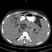
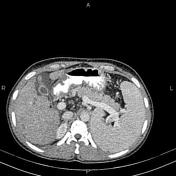
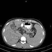
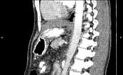
Mild to moderate free fluid is noted in abdominopelvic spaces.
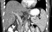
The liver has irregular margin is keeping with cirrhosis. Numerous small ill-defined low- enhancing masses are noted throughout the liver, most consistent with regenerative nodules. Intra- and extrahepatic bile ducts are of normal caliber.
The gallbladder shows diffuse wall thickening without CT-detectable stone.
The spleen is enlarged and its cephalocaudal height measured 145mm.
Mild degenerative changes as osteophytosis are seen at the lumbar spine.
Case Discussion
Features on CT scan consistent with cirrhosis with secondary splenomegaly.


 Unable to process the form. Check for errors and try again.