
Presentation
Previous history of motor vehicle trauma.
Patient Data


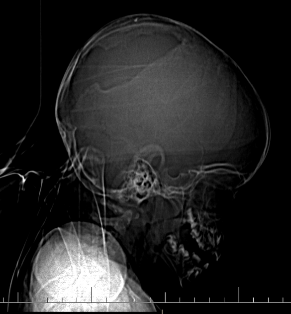
Oval shape lucency with smooth margins at parietal bone is detected in scanogram.
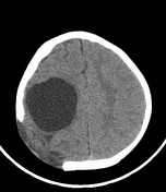
A lytic calvarial lesion with scalloped edges at right parietal bone is visible with extracranial brain herniation. A large porencephalic cyst at right frontoparietal lobe is detected. Right sided unilateral ventricular dilatation is seen.
Based on previous history of trauma and the above-mentioned findings, leptomeningeal cyst or growing fracture should be considered.
Case Discussion
CT scan is the modality of choice for the evaluation of leptomeningeal cyst. It consists of a lytic calvarial lesion with scalloped edges, in which encephalomalacia invaginates. The following features may also be present 1,2:
extracranial brain herniation
hydrocephalus
unilateral ventricular dilatation
porencephalic cyst


 Unable to process the form. Check for errors and try again.