
Presentation
Abdominal pain.
Patient Data
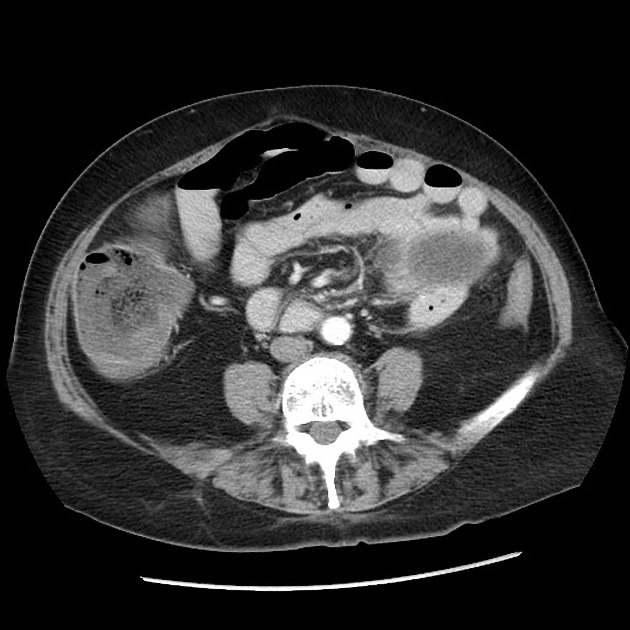
Colonic obstruction with transition point at the splenic flexure with a narrowed, curved, angulated segment surrounded by omental fat. No mass identified.
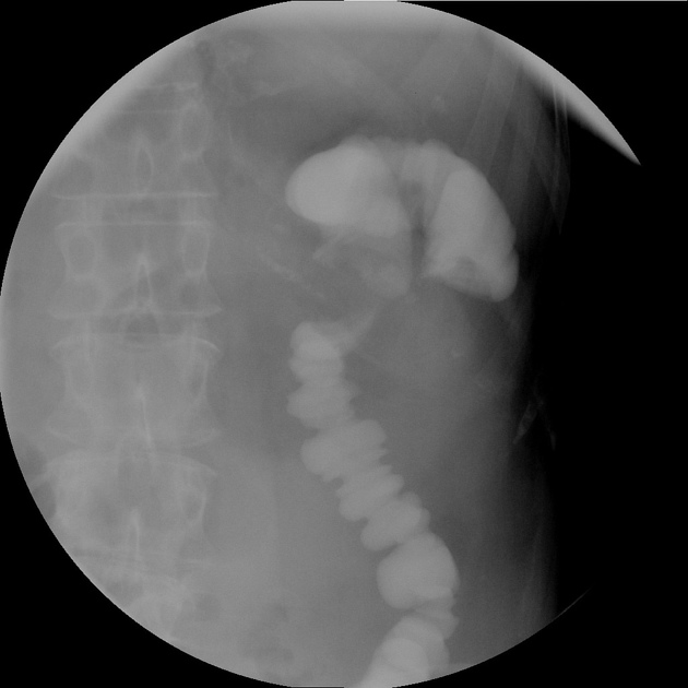
Gastrografin enema with retrograde filling of the descending colon to the splenic flexure, with angulation and narrowing with failure to fill into the transverse colon indicating obstruction.
Case Discussion
An uncommon type of internal hernia caused by transomental herniation of the splenic flexure, with some component of adhesions based on the operative note. The angulated, narrowed appearance of the bowel indicates some degree of internal herniation and/or adhesions causing this obstruction. Gastrografin enema was performed preoperatively which confirmed colonic obstruction as no contrast passed retrograde beyond the splenic flexure.
Operative note (edited excerpt): "... A laparoscope was placed through the port and attention turned to the left upper quadrant where we identified an internal hernia of the transverse colon just proximal to the splenic flexure. The colon appeared viable but was caught in the omentum of the transverse colon. A decision was made to attempt to reduce this laparoscopically and several more laparoscopic ports were placed...using atraumatic graspers, we were able to identify an internal hernia equivalent caused by adhesions of the omentum. We were able to free up a portion of the omentum using the harmonic scalpel and divide it. After the division of the omentum, we were able to reduce the hernia..."


 Unable to process the form. Check for errors and try again.