
Presentation
4 days of abdominal pain, nausea and vomiting with constipation.
Patient Data
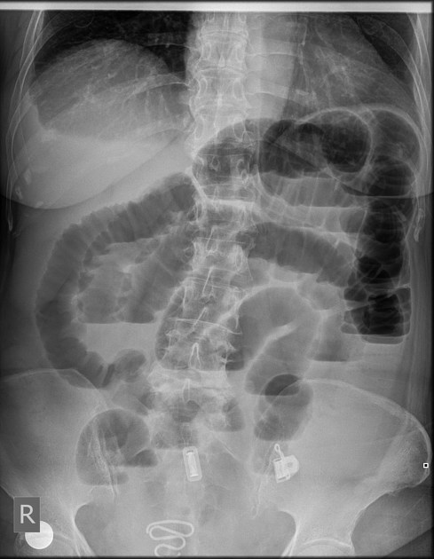
Erect plain abdomen x-ray shows:
- dilated small bowel loops with multiple gas-fluid levels
- there is a pelvic metallic coil consistent with an IUCD
- the right C/P angle is seen obliterated suggesting mild pleural effusion
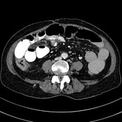

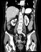
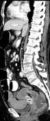
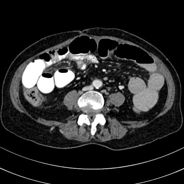
Small bowel long segment abnormal dilatation; transition zone of high grade caliber change is seen at the right adnexa where there is a short segment of closed loop obstruction of the distal ileum with its neck seen just related to the right fallopian tube/right uterine vessels. The terminal ileum and the rest of the bowel loops located distally to the transition zone are seen collapsed with an empty rectum.
There are engorged mesenteric vessels with noted abdomen and pelvic free fluid suggesting mesenteric congestion.
No radiologic evidence of bowel ischemia or perforation.
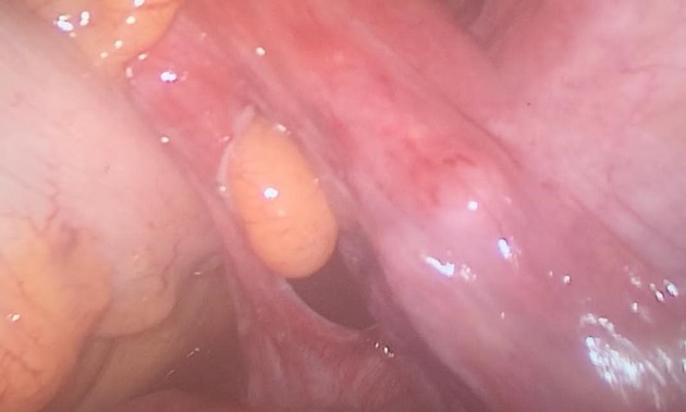
Image by the operative laparoscopy showing the broad ligament defect (right side) after releasing the herniated bowel loops
Case Discussion
The CT findings are in keeping with complete bowel obstruction secondary to a broad ligament internal hernia (of the right side).
The patient was operated laparoscopically, a herniated short segment of the terminal ileum through a broad ligament defect was found. The herniated bowel loop was viable and clean with no evidence of incarceration. The broad ligament defect was closed with surgical sutures.
The patient discharged after 2 days with no postoperative complications.


 Unable to process the form. Check for errors and try again.