
Presentation
Chest pain Shortness of breath
Patient Data

Heart is enlarged . CTR is 0.6
Blunting of right costophrenic angle suggestive of pleural effusion.
Fluid extending to right minor fissure.
Median sternotomy wires in situ with mediastinal surgical clips consistent with previous CABG
There is upper lobe venous diversion.
Coarsening of bronchovascular markings suggestive of COPD.
Air space opacities at both lung bases more on left
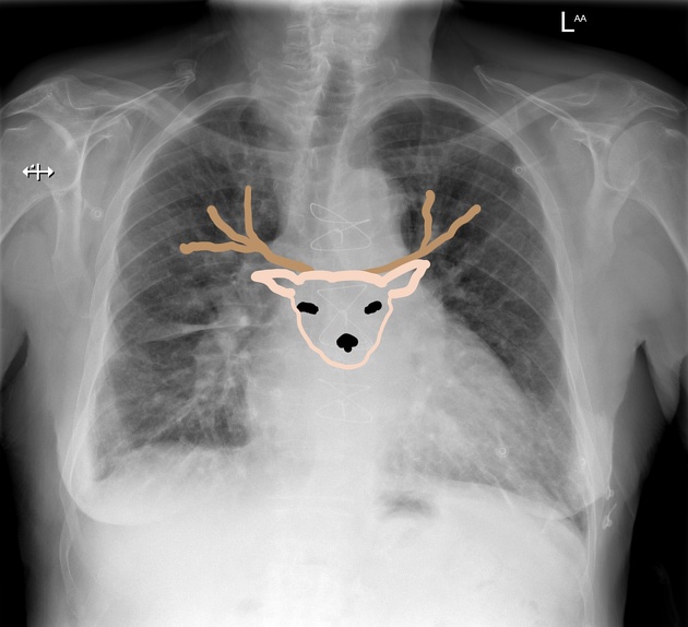
Dilated and branching upper zone pulmonary veins resemble the antlers of a stag
Case Discussion
Pulmonary edema is graded depending on chest x-ray and pulmonary capillary wedge pressure (PCWP) is as follows:
- Grade 0: normal chest radiograph, PCWP 8-12 mmHg
- Grade 1: upper lobe diversion on a chest radiograph, PCWP 13-18 mmHg
- Grade 2: interstitial edema on a chest radiograph, PCWP 19-25 mmHg
- Grade 3: alveolar edema on a chest radiograph, PCWP >25 mmHg
Our case is representing grade 1 pulmonary edema.
Upper lobe pulmonary venous diversion (cephalisation) reflects elevation of left atrial pressure and can occur with pulmonary edema
Stag antler sign: The prominence of upper lobe pulmonary veins resembles a stag's antlers. It is the earliest sign of pulmonary venous hypertension (grade 1 pulmonary edema).


 Unable to process the form. Check for errors and try again.