
Patient Data
Age: 40 years
Gender: Female

Download
Info
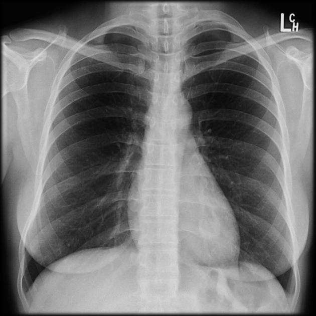
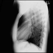
Chest x-ray demonstrates a founded well-circumscribed lesion projecting through the cardiac silhouette on the left. On lateral projection, it is visible abutting the heart posteriorly.


Download
Info
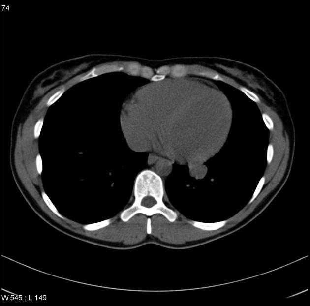
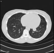
CT confirms the presence of the lesion which is of fluid attenuation. It is spherical in shape abutting the posterior aspect of the heart appearing to be within the left lung base.
Download
Info

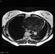
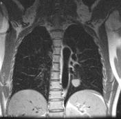
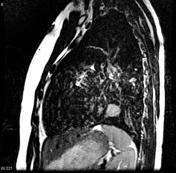
MRI demonstrates the lesion to be of high T2 signal. It is spherical in shape abutting the posterior aspect of the heart appearing to be within the left lung base.
Case Discussion
Appearances of this mediastinal cyst most likely represent a bronchogenic cyst, with the alternative diagnosis being that of a pericardial cyst. Unfortunately, no followup or histology is available.


 Unable to process the form. Check for errors and try again.