
Presentation
Pelvic pain and mass feeling.
Patient Data
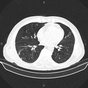


Patchy ill-defined subpleural ground glass opacities are seen at both lung bases suggestive for COVID-19 pneumonia.
Degenerative changes as osteophytosis are seen at the thoracic spine.
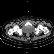
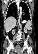
Several small non-enhancing cysts are seen at liver.
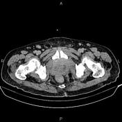
Several non-enhancing simple cortical cysts are seen at both kidneys, with maximum diameters of 55 mm.
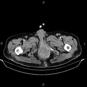
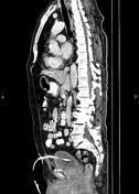
An 85×60×65 mm heterogeneously enhancing mass is present at lower pelvis, inferior to the urinary bladder that infiltrates vesical base, prostate gland, rectum and perirectal / perianal regions. Several lymphadenopathies are seen in the vicinity of diseased segment with maximum SAD of 14 mm. Suprapubic catheter is inserted into the urinary bladder.
Advanced degenerative changes as osteophytosis are seen at the lumbar spine.
Grade I spondylolisthesis of L5 on S1 is present with bilateral spondylolysis.
Case Discussion
Pelvic mass with local invasion; pathology proven prostate adenocarcinoma; with regional lymphadenopathies.
Prostate cancer can spread by local invasion (typically into the bladder and seminal vesicles; urethral and rectal involvement are rare), lymphatic spread (pelvic nodes first followed by para-aortic and inguinal nodes), or by hematogenous metastases.


 Unable to process the form. Check for errors and try again.