
Presentation
Central abdominal pain for the last 2 days.
Patient Data
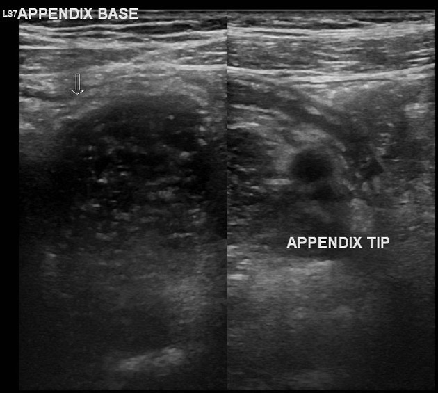
The appendix was evaluated in its entire length. It is in the right iliac fossa. The length of the appendix was about 90 mm. The proximal about 40 mm length of the appendix shows normal diameter (< 6 mm), compressibility. The distal part of the appendix shows dilatation (diameter > 6 mm), non-compressibility, increased vascularity, intact non-distended lumen with preserve gut signature sign. There is no fecolith at the site of transition from dilated to a normal appendix. There is periappendiceal fat inflammation around the distal appendix. The cecum and terminal ileum were normal.
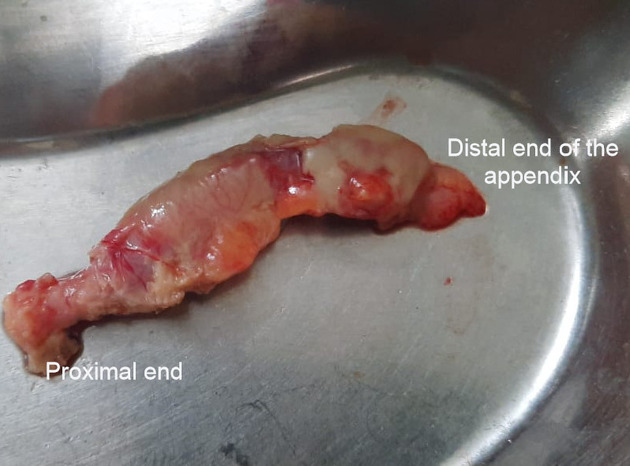
The resected appendix shows normal diameter of the proximal segment. The rest of the appendix is dilated. There is no wall perforation.
Case Discussion
The case shows the importance of the scanning entire length of the appendix to rule out/ rule in acute appendicitis.
Gross specimen photo courtesy: operatingng surgeon Dr. Niraj I. Patel.


 Unable to process the form. Check for errors and try again.