
Presentation
Small painless stable swelling in the left lower posterior chest wall since birth. Now the patient presented with pain in the swelling after trauma to the left chest wall. No discharge or fever.
Patient Data

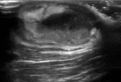
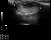

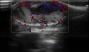
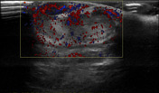
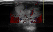
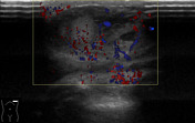

Scan demonstrates a well-defined heterogeneous mixed echogenicity lesion measuring approximately 30 x 17 mm in the superficial subcutaneous soft tissues of the left lower posterior chest wall. Moderate internal vascularity is seen in it on color Doppler ultrasound examination, particularly along its periphery. No definite connection is appreciable with the underlying muscles or chest cavity.
Case Discussion
Based on the history of soft tissue swelling since birth and the above mentioned sonographic features, diagnosis of a low-grade soft tissue tumor-like dermatofibrosarcoma protuberans was given. Because of history of recent trauma, imaging differentials of hematoma with superadded infection, or hemorrhage/infection in a pre-existing sebaceous cyst or dermoid cyst were given which were however unlikely.
Pathology
Procedure: Excision of left lower posterior chest wall skin lesion.
Diagnosis: Dermatofibrosarcoma protuberans (DFSP). Greatest dimension of tumor: 3 cm. Extent of tumor: Dermal & subcutaneous tissue. Mitotic rate: One/10 high power fields. Necrosis: Not identified. Margins: Clear. Lymphovascular invasion: Not identified. Immunohistochemistry: CD34 and vimentin are diffusely and strongly positive (with internal positive control). S100 and cytokeratin AE1/AE3 are both negative (with positive internal control).


 Unable to process the form. Check for errors and try again.