
Presentation
Right flank pain.
Patient Data
Age: 60 years
Gender: Male


Download
Info
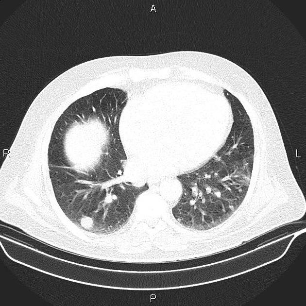
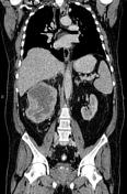
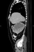
Multiple intrapulmonary nodules are seen inferring metastasis.
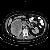
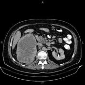
A large hetero enhancing mass is seen at right kidney accompanied by perirenal fat infiltration and multiple dilated perinephric collateral vessels. The mass is extended to right renal vein but there is no sign of extension to IVC.
The prostate gland is enlarged.
From the case:
Renal cell carcinoma
Download
Info
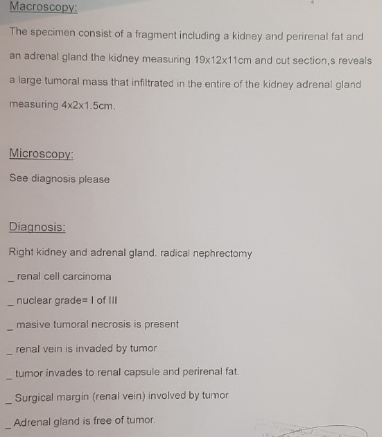
Right nephrectomy and adrenalectomy performed for the patient and histopathology evaluation confirms renal cell carcinoma with vascular extension.


 Unable to process the form. Check for errors and try again.