
Presentation
Concern of abdominal wall hernia.
Patient Data
Age: 30 years
Gender: Male
From the case:
Omental solitary fibrous tumor

Download
Info

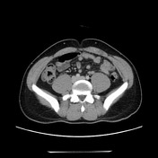
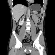
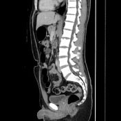
Lobulated enhancing omental nodule left of the umbilicus, 1.5 x 2.0 cm. No other acute findings.
Case Discussion
Incidental finding of an enhancing omental nodule, which underwent ultrasound guided biopsy and ultimately surgical resection. Final pathology revealed "Solitary fibrous tumor with intact capsule; no evidence of tumor necrosis and low mitotic count".


 Unable to process the form. Check for errors and try again.