
Presentation
33/40 pregnant. Acute right-sided abdominal pain and pyrexia.
Patient Data


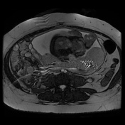
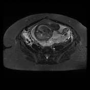
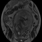
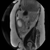
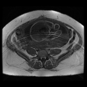
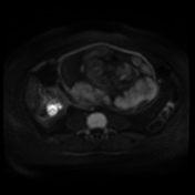
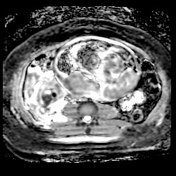
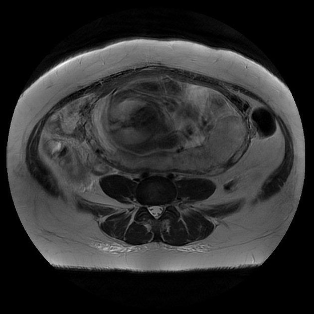
Intrauterine pregnancy confirmed but not interrogated in detail. The gravid uterus displaces the cecal pole and appendix out of the right iliac fossa. An acute inflammatory mass is present adjacent to the cecal pole, medially and posteriorly, with edema of the fat and a small volume of free fluid. Appendicoliths are seen within the distended edematous appendix, with a small fluid collection immediately adjacent. Appearances consistent with acute appendicitis with localized perforation and fluid collection. Incidental note is made of distension of the right ovarian vein.
Histopathology
Clinical Details: Perforated appendix.
Macroscopic: Perforated appendix 61x10mm with fibrinopurulent exudate on the surface.
Microscopic: This appendix shows severe transmural active inflammation with extensive tissue necrosis extending to the serosal layer, consistent with a perforation. There is no evidence of worms, granulomas, dysplasia or malignancy.
Conclusion: Appendix - perforated acute gangrenous appendicitis.
Case Discussion
The MRI shows signs of complicated acute appendicitis with a phlebolith in the lumen of the thickened appendix and a fluid collection adjacent and generalized edema of the surrounding tissue planes. This study also demonstrated an engorged right ovarian vein, seen here as high signal on the gradient echo images, but predominantly low signal on the SSFSE image, due to flow voids.


 Unable to process the form. Check for errors and try again.