
Presentation
21/40 pregnant. Acute right-sided abominal pain.
Patient Data
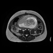

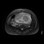
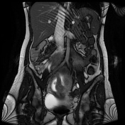

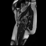

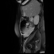
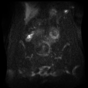
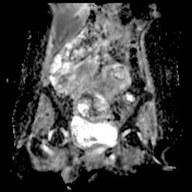
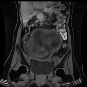
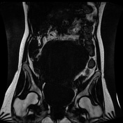

Intrauterine pregnancy confirmed but not interrogated in detail. The gravid uterus displaces the cecal pole and appendix out of the right iliac fossa. An acute inflammatory mass is present adjacent to the cecal pole, medially and posteriorly, with edema of the fat and a small volume of free fluid. Appendicoliths are seen within the distended edematous appendix. There is restricted diffusion. The appearances are consistent with acute appendicitis. The right ovary, containing a small peripheral cyst, is located very close to the inflammatory appendix mass.
Incidental note is made of right-sided physiologic hydronephrosis caused by compression of the ureter between the uterus and the right psoas muscle, and distension of the right ovarian vein.
Histopathology
Clinical details: Appendicitis
Macroscopic: Swollen appendix 15 x 12 x 10 mm with exudate on surface. Defect on surface ?Perforation, 10 mm in length.
Microscopic: Sections taken from the appendix show foreign transmural acute inflammation with necrosis of the wall. The defect seen macroscopically comprises an area showing fecolith with gangrenous changes in the wall. There are some areas in which the continuity of the wall appears interrupted, suggestive of perforation.
Conclusion: Appendix - gangrenous appendicitis
Case Discussion
The MRI shows signs of uncomplicated acute appendicitis with phleboliths in the lumen of the thickened appendix and generalized edema of the surrounding tissue planes. The appendix lies very close to the right ovary and care must be taken to interrogate the region of the cecal pole carefully for signs of inflammation on fat-saturated and diffusion images, and to look for luminal appendiceal distension with appendicoliths.
This study also demonstrated two tubular structures in the right retroperitoneum that may be mistaken for each other, or for the appendix; the engorged right ovarian vein and the distended right ureter. Both are seen on the gradient echo images as high signal tubular structures, the signal of the ovarian vein may be altered in the presence of ovarian vein thrombosis.


 Unable to process the form. Check for errors and try again.