
Presentation
22/40 pregnant. Acute right-sided abdominal pain.
Patient Data


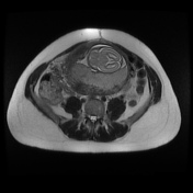

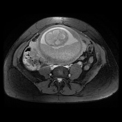

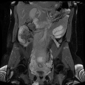
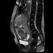
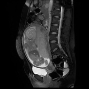
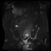
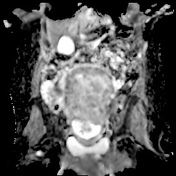
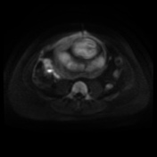
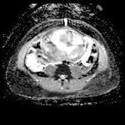
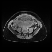


The gravid uterus displaces the cecal pole out of the right iliac fossa. The right ovary lies close to the cecal pole, and the appendix is not identified confidently as a separate structure, but the diffusion-weighted imaging indicated an inflamed curvilinear structure close to the ovary and cecal pole, consistent with acute appendicitis. Incidental note is made of distension of the right ovarian vein.
Histopathology
Clinical Details: Appendectomy
Macroscopic: Appendix 47 x 12 mm with fibrinopurulent exudate on surface
Microscopic: This is acute suppurative appendicitis with peritonitis. There is no evidence of neoplasia and no parasites are seen.
Conclusion: Acute suppurative appendicitis
Case Discussion
The appendix may not be clearly identified on MRI in pregnancy for a number of reasons, including the limited spatial resolution compared to CT, and the alteration in anatomy caused by the gravid uterus. In this case, although not identified as a distinct structure due to the proximity of the structures in the right side of the abdomen, the signal change on the diffusion-weighted imaging provide the clue to the presence of inflammatory change. Acute appendicitis was confirmed at pathology analysis.
This study also demonstrated an engorged right ovarian vein, seen here as high signal on the gradient echo images, but predominantly low signal on the SSFSE image, due to flow voids.


 Unable to process the form. Check for errors and try again.