
Presentation
Abdominal pain and dyspepsia
Patient Data
Age: 50 years
Gender: Female
From the case:
Gastric adenocarcinoma

Download
Info

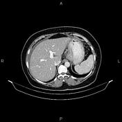
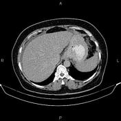
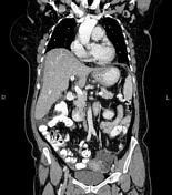
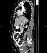
Increased wall thickness due to tumoral infiltration is present at gastric cardia and proximal of lesser curvature accompanied by a few small regional lymphadenopathies. Mild abdominopelvic ascites with omental thickening also evident.
An IUCD is present within the uterine cavity in appropriate location.
Case Discussion
Gastric mass (pathology proven adenocarcinoma) with regional lymphadenopathies; ascites and peritoneal seeding.
CT is currently the staging modality of choice because it can help identify the primary tumor, assess for the local spread, and detect nodal involvement and distant metastases.


 Unable to process the form. Check for errors and try again.