
Patient Data
Age: Young adult
Gender: Female
From the case:
Cavernous malformation with DVA


Download
Info

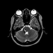
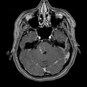
MRI demonstrates a right pontine popcorn lesion with a complete surrounding hemosiderin ring with complex signal intensity due to blood product of varying age. The lesion is consistent with a cavernous malformation. Note the presence of an associated developmental venous anomaly seen only on T1 C+ sequence, draining the right cerebellar hemisphere. Susceptibility sequence failed to detect other similar lesions.
Case Discussion
Typical appearances of a cavernous malformation (Zabramski type 2) with an associated DVA, a very common finding.


 Unable to process the form. Check for errors and try again.