
Presentation
Incidental finding. Work up for abdominal pain.
Patient Data
Age: 45 years
Gender: Female
From the case:
Renal cell carcinoma

Download
Info

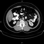
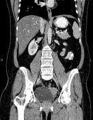
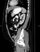
A 22 mm exophytic mass (86HU in attenuation value) was seen in the lower pole of the left kidney.
Case Discussion
Ultrasonography evaluation shows that the left renal lesion is a solid mass and not a complicated cyst. The patient underwent a left partial nephrectomy, and histopathology confirmed a renal cell carcinoma, the most common malignant renal tumor.


 Unable to process the form. Check for errors and try again.