
Presentation
Known seizure disorder. Increasing headaches and seizure frequency.
Patient Data
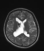

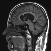
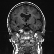
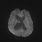
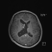

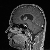

An enhancing mixed solid/cystic lesion contacts the lateral wall of the right lateral ventricle and the right foramen of Monro. Assymetric enlargement of the right lateral ventricle with leftward bowing of the left lateral ventricle.
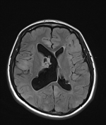
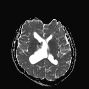
A smaller non-enhancing nodular lesion arises from the posterior body of the left ventricle/left trigone.
There are multiple cortical and subcortical regions of FLAIR hyperintensity without enhancement, reflecting cortical tubers. Areas of low T1 and T2 are likely to represent calcified tubers.
Case Discussion
In a patient with tuberous sclerosis, an enhancing lesion at the foramen of Monro is typical of SEGA. Distinguishing between an enhancing subependymal nodule is based on growth over interval studies.


 Unable to process the form. Check for errors and try again.