Presentation
Abdominal pain (lumbar region) for 10 days, associated with a few episodes of vomiting. No dysuria or fever.
Patient Data
![](https://prod-images-static.radiopaedia.org/images/54956254/3_thumb.jpeg)
![](https://prod-images-static.radiopaedia.org/images/54956252/1_thumb.jpeg)
![](https://prod-images-static.radiopaedia.org/images/54956258/7_thumb.jpeg)
![](https://prod-images-static.radiopaedia.org/images/54956257/6_thumb.jpeg)
![](https://prod-images-static.radiopaedia.org/images/54956255/5_-_Copy_thumb.jpeg)
![](https://prod-images-static.radiopaedia.org/images/54956260/9_thumb.jpeg)
![](https://prod-images-static.radiopaedia.org/images/54956254/3_big_gallery.jpeg)
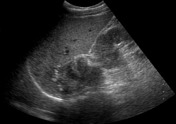
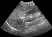
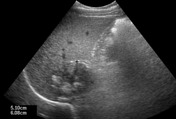
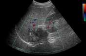
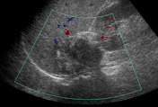
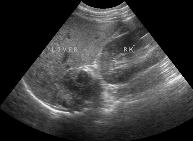
Heterogeneous partly calcified mass lesion measuring 5 x 6 cm, in right suprarenal location. No vascularity is seen in it on color Doppler ultrasound examination.
![](https://prod-images-static.radiopaedia.org/images/54956490/Anonymize.Seq2.Ser2.Img149_thumb.jpeg)
![This study is a stack](/packs/stack-YQKLCKBI.gif)
![](https://prod-images-static.radiopaedia.org/images/54956320/Anonymize.Seq3.Ser3.Img117_thumb.jpeg)
![This study is a stack](/packs/stack-YQKLCKBI.gif)
![](https://prod-images-static.radiopaedia.org/images/54956781/Anonymize.Seq3.Ser3.Img845_thumb.jpeg)
![This study is a stack](/packs/stack-YQKLCKBI.gif)
![](https://prod-images-static.radiopaedia.org/images/54957288/Anonymize.Seq4.Ser200.Img37_thumb.jpeg)
![This study is a stack](/packs/stack-YQKLCKBI.gif)
![](https://prod-images-static.radiopaedia.org/images/54956639/Anonymize.Seq7.Ser301.Img35_thumb.jpeg)
![This study is a stack](/packs/stack-YQKLCKBI.gif)
![](https://prod-images-static.radiopaedia.org/images/54957430/Anonymize.Seq7.Ser301.Img101_thumb.jpeg)
![This study is a stack](/packs/stack-YQKLCKBI.gif)
![](https://prod-images-static.radiopaedia.org/images/54957640/Anonymize.Seq5.Ser201.Img39_thumb.jpeg)
![This study is a stack](/packs/stack-YQKLCKBI.gif)
![](https://prod-images-static.radiopaedia.org/images/54957492/Anonymize.Seq6.Ser300.Img39_thumb.jpeg)
![This study is a stack](/packs/stack-YQKLCKBI.gif)
![](https://prod-images-static.radiopaedia.org/images/54957565/Anonymize.Seq6.Ser300.Img185_thumb.jpeg)
![This study is a stack](/packs/stack-YQKLCKBI.gif)
![](https://prod-images-static.radiopaedia.org/images/54956490/Anonymize.Seq2.Ser2.Img149_big_gallery.jpeg)
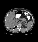
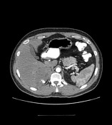
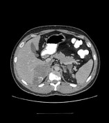
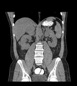
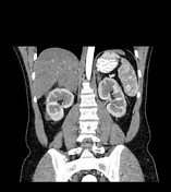
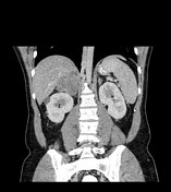
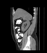
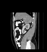
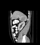
Right suprarenal mass lesion measuring about 5.7 x 5.4 cm, containing multiple flocculent calcifications, showing enhancement of the soft tissue components in the post-contrast study. Partially thrombosed inferior vena cava. Multiple para-aortic and renal hilar lymph nodes, with the largest one measuring 1.2 cm in short axis. Multiple simple and a few hyperdense (hemorrhagic/complicated) bilateral renal cysts.
![](https://prod-images-static.radiopaedia.org/images/54959988/ganglioneuroma-8__1__thumb.jpeg)
![](https://prod-images-static.radiopaedia.org/images/54960051/ganglioneuroma-8__2__thumb.jpeg)
![](https://prod-images-static.radiopaedia.org/images/54957699/Coronal_thumb.jpeg)
![](https://prod-images-static.radiopaedia.org/images/54959988/ganglioneuroma-8__1__big_gallery.jpeg)
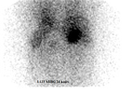
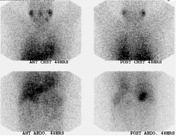
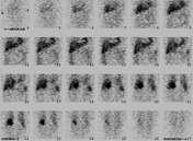
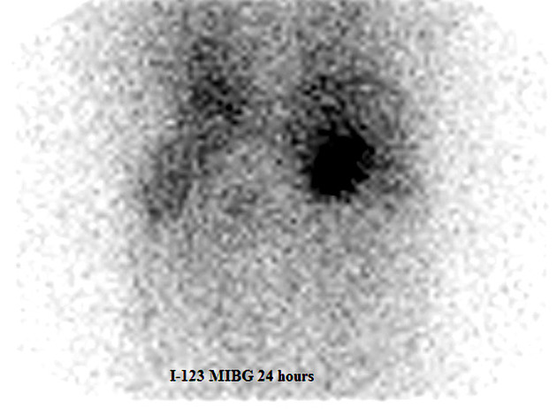
Abnormal I-123 MIBG uptake in the right suprarenal region, with no evidence of MIBG-avid disease elsewhere.
Case Discussion
Procedure: Open right adrenalectomy.
Diagnosis: Ganglioneuroma (schwannian stroma-dominant).
Immunohistochemistry: S100 (polyclonal) is strongly positive. SMA (Alpha-SM1) is negative.
40 % of ganglioneuromas are seen in patients over the age of 20 years and 20 % are within the adrenal gland.