
Presentation
Pelvic pain.
Patient Data
Age: 50 years
Gender: Female
From the case:
Hemangiopericytoma - pelvis

Download
Info

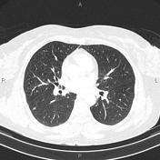
A few subpleural and intraparenchymal nodules less than 5mm in diameter are seen at both lungs.
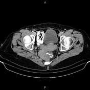
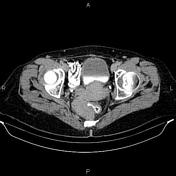
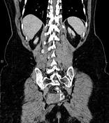
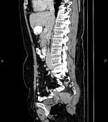
A 68×35mm well-defined soft tissue mass is seen at right distal presacral region. Fat plane between the mass and adjacent rectal wall is obliterated.
Case Discussion
Pathology proven hemangiopericytoma, which is a term formerly used to describe a continuum of mesenchymal tumors with elevated cellularity found throughout the body in soft tissue and bone.


 Unable to process the form. Check for errors and try again.